
Fig. 3.
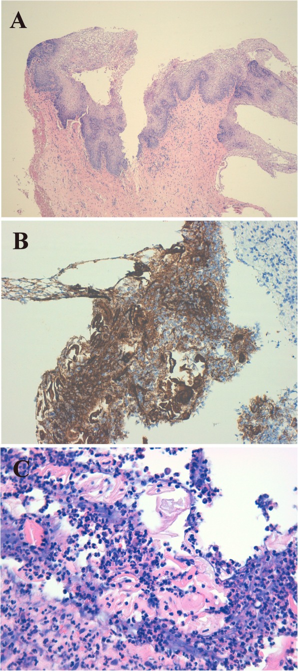
Vaginal biopsy at 6 months. a Well-structured, mature squamous epithelium (haematoxylin-eosin [H-E], × 40) in c4. b Intense staining for cytokeratin AE1-AE3 (CKAE1-AE3) on the surface of the fragment, which helps in recognizing the epithelium at that level and with greater magnification to see the epithelial positivity to cytokeratins (CKAE1-AE3, × 200). c Among abundant inflammatory polymorphic cells, the presence of keratin sheets (dyed more homogeneous pink) can be estimated. With greater magnification, the keratin sheets are more evident (H-E, × 400)