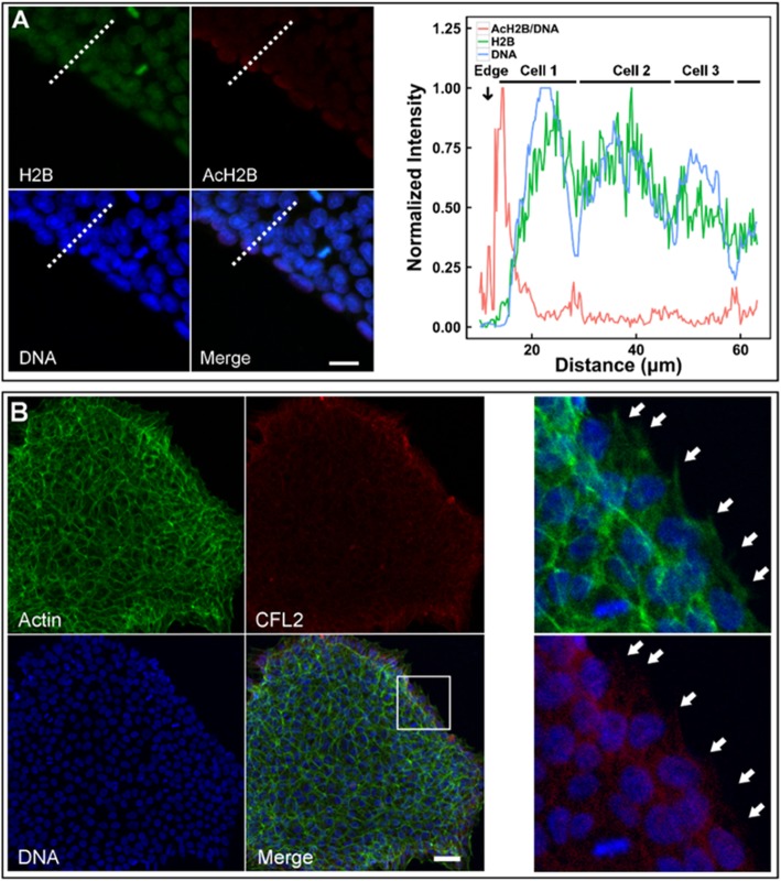
Fig. 4.
H2B acetylation and CFL2/F-actin cytoskeleton at the edge of a colony. Expressions of AcH2B/DNA, DNA, and AcH2B were quantified (a; red, blue, and green lines in right panel denotes, respectively, AcH2B/DNA, DNA, and H2B expressions). Also plotted were the distributed F-actin and CFL2 proteins at the edge of a typical colony (b). All intensity values were normalized to (0, 1) upon the formulation of <Xi > = (Xi − Xmin)/(Xmax − Xmin), where Xi = data point i and Xmin or Xmax = the minimum or maximum value among all the data points. Solid arrows in b indicated the pseudopodia. Bar = 20 μm in a and 50 μm in b

