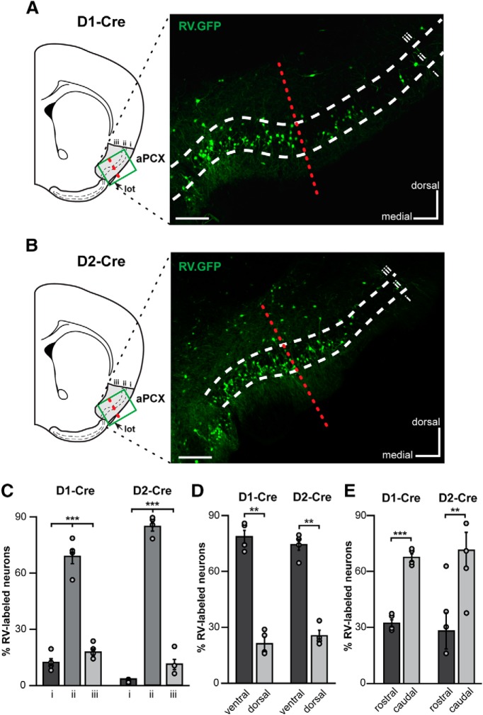
Figure 8.
Neurons from the ventrocaudal aPCX innervate OT D1- and D2- type MSNs. A, B, Coronal brain section schematic of analyses regions (∼0 mm A/P bregma) and representative coronal brain sections displaying aPCX neurons labeled by the rabies virus (RV) (RV-EnvA-ΔG-GFP, green) from the OT in both D1-Cre (A) and D2-Cre mice (B). Scale bar, 200 μm. Green boxes in schematics indicate approximate regions used to collect images for analyses. Red dashed line indicates the approximate boundary of ventral versus dorsal aPCX. C, Percentage of RV+ neurons across all aPCX layers. D, Percentage of RV+ neurons localized within the ventral and dorsal aPCX subregions. E, Percentage of RV+ neurons localized within the rostral and caudal aPCX subregions. Individual data points in C to E indicate within animal means. **p < 0.01, ***p < 0.0001.

