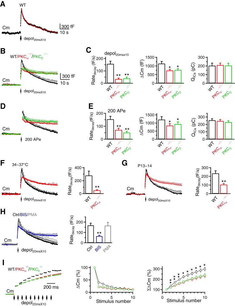
Figure 3.
PKCα or PKCβ knock-out inhibits rapid endocytosis and vesicle mobilization to the readily releasable pool at calyces. A–H, Similar arrangements as Figure 2A–H, respectively, except that the stimulus was depol20msX10 (A–C, F–H) or 200 APe at 100 Hz (D,E) for inducing rapid endocytosis. A, Red curve is a biexponential fit of the Cm decay with a τ of 1.4 s and 14.3 s, respectively (ICa not shown). B, C, WT, 9c/9m; PKCα−/−, 14c/14m; PKCβ−/−, 6c/6m. D, E, WT, 8c/8m; PKCα−/−, 11c/11m; PKCβ−/−, 8c/8m. F, WT, 6c/6m; PKCα−/−, 4c/4m. G, WT, 7c/7m; PKCα−/−, 8c/8m. H, Ctrl, 11c/11m; BIS, 11c/11m; PMA, 8c/8m. I, Left, Sampled Cm induced by depol20msX10 (each arrow: 1 depol20ms) from WT, PKCα−/− and PKCβ−/− calyces. ΔCm induced by the first depol20ms was normalized. Middle and right, ΔCm (middle) and accumulated ΔCm (ΣΔCm, right) induced by each of the 10 depol20ms during depol20msX10 in WT (9c/9m), PKCα−/− (14c/14m) and PKCβ−/− (6c/6m) calyces. Data (mean ± SEM) are normalized to ΔCm induced by the first depol20ms. ΣΔCm was significantly higher for the WT group (*p < 0.05; **p < 0.01).

