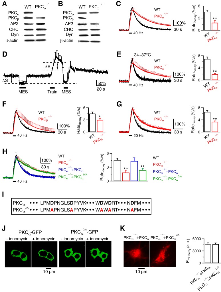
Figure 4.
PKC and its calcium-binding domain are required for endocytosis at hippocampal synapses. A, B, Western blot of PKCα, PKCβ, adaptor protein 2 (AP2), clathrin heavy chain (CHC), dynamin (Dyn), and β-actin in WT, PKCα−/− (A), and PKCβ−/− (B) hippocampal culture. Results in A and B were repeated by 2–4 times. C, FSypH traces (normalized to baseline, left) and Ratedecay (right) induced by Train40Hz (bar) in WT (n = 14 experiments) or PKCα−/− (n = 28 experiments) hippocampal culture at 22–24°C. Data plotted as mean + SEM; *p < 0.05; **p < 0.01, t test (applies to all similar graphs). Throughout the study, each experiment contained 20–30 boutons; 1–3 experiments were taken from 1 culture; each culture was from 3–5 mice; each group was from 4–12 cultures. D, Applying MES solution (pH:5.5, bars) quenched FSypH (mean + SEM) to a similar level (lowest dash line) before and after a 10 s train of stimuli in PKCα−/− boutons (n = 6 experiments, 22–24°C). ΔS, SypH at resting plasma membrane quenched by MES. E–G, Similar to C, but at 34–37°C (E), in PKCβ−/− culture (F), or after a 10 s train at 20 Hz (G). E, WT, n = 6 experiments; PKCα−/−, n = 6. F, WT, n = 14; PKCβ−/−, n = 5. G, WT, n = 16; PKCα−/−, n = 22. H, FSypH traces and Ratedecay induced by Train40Hz (bar) in WT boutons (n = 14), PKCα−/− boutons (PKCα−/−, n = 28), PKCα−/− boutons rescued with WT PKCα (containing mCherry for recognition, PKCα−/−+PKCα, n = 7), and in PKCα−/− boutons rescued with PKCαD/A and mCherry (PKCα−/−+PKCαD/A, n = 8). I, Protein sequence of PKCα and PKCαD/A C2 domain. The Ca2+-coordinating aspartates of PKCα (bold) were mutated to alanines (red) in PKCαD/A. J, We expressed PKCα-GFP (left two panels) or PKCαD/A-GFP (right two panels) in HEK293T cells and monitored the subcellular distribution of the kinase. The Ca2+ ionophore, ionomycin (10 μm, 15 min), induced translocation of PKCα-GFP toward the plasma membrane, but did not alter the intracellular distribution of PKCαD/A-GFP. Such results were observed in 3 experiments (each experiment had 2–3 cells). K, Left, PKCα−/− neurons rescued with WT PKCα (containing mCherry for recognition, PKCα−/−+PKCα), or with PKCαD/A and mCherry (PKCα−/−+PKCαD/A). Right, Fluorescence intensity of mCherry (FmCherry) in PKCα−/−+PKCα neurons (n = 10) and PKCα−/−+PKCαD/A neurons (n = 13). FmCherry was measured from both soma and branches.

