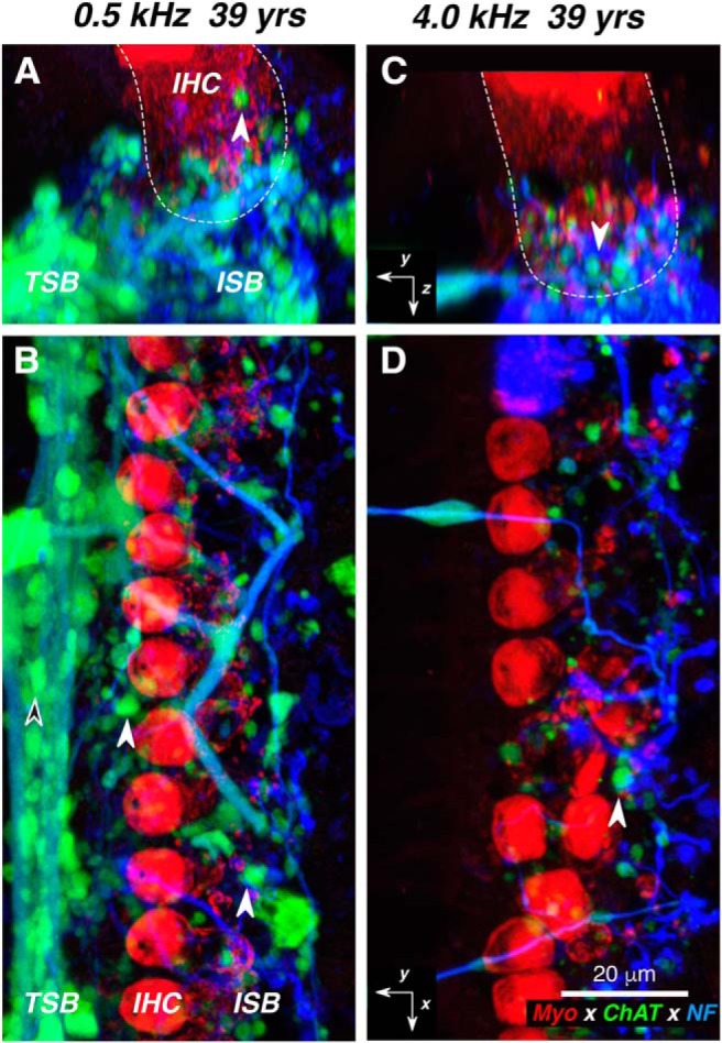
Figure 6.

A–D, Confocal images of the LOC innervation of IHCs from the apical (A, B) vs basal (C, D) half of the cochlea from one subject. At each locus, two views of the same confocal z-stack are shown: B and D are maximum projections in the acquisition plane (xy), while A and C are maximum projections of the entire stacks in the zy plane, with the approximate boundaries of the IHCs indicated in white dashed lines. White-filled arrowhead in all panels point to ChAT-positive boutons in the ISB, while the black-filled arrowhead in B points to a ChAT-positive bouton in the TSB. Immunostaining key and scale bar in D apply to all panels, and the orientation arrows in C and D also apply to A and B, respectively: the x arrows point along the spiral toward the apex, y arrows point radially away from the modiolus, and z arrows point toward scala tympani. yrs, Years; NF, neurofilament.
