Figure 2.
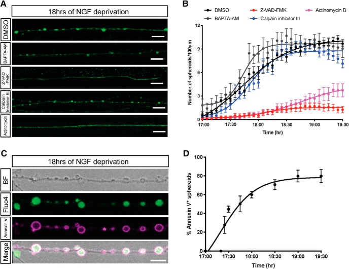
Transcription and caspase activation are required for formation of spheroids. A, B, Fluo4-AM calcium imaging (A) and quantification of axonal spheroid number per 100 μm of sympathetic axons (B) at the indicated times after NGF deprivation in the presence of DMSO, 10 μm BAPTA-AM, 50 μm V-ZAD-FMK, 20 μm calpain inhibitor III, and 1 μg/ml actinomycin, respectively. Scale bar, 10 μm. Nonlinear regression curves were drawn according to the Hill equation. Total number of n = 26 (DMSO), n = 26 (BAPTA-AM), n = 25 (Z-VAD-FMK), n = 20 (calpain inhibitor III), and n = 17 (actinomycin) axons from three independent litters were counted. C, Representative images of axonal spheroids after 18 h of NGF deprivation. Fluo4-AM (green) indicates intra-axonal calcium, and annexin V (magenta) indicates exposure of PS on the outer leaflet on the spheroid membrane. Scale bar, 5 μm. D, Quantification of the percentage of fluorescent annexin V-positive spheroids after 17–19.5 h of NGF deprivation. Total number of n = 15 axons from three independent litters were counted. Data are mean ± SEM.

