Figure 4.
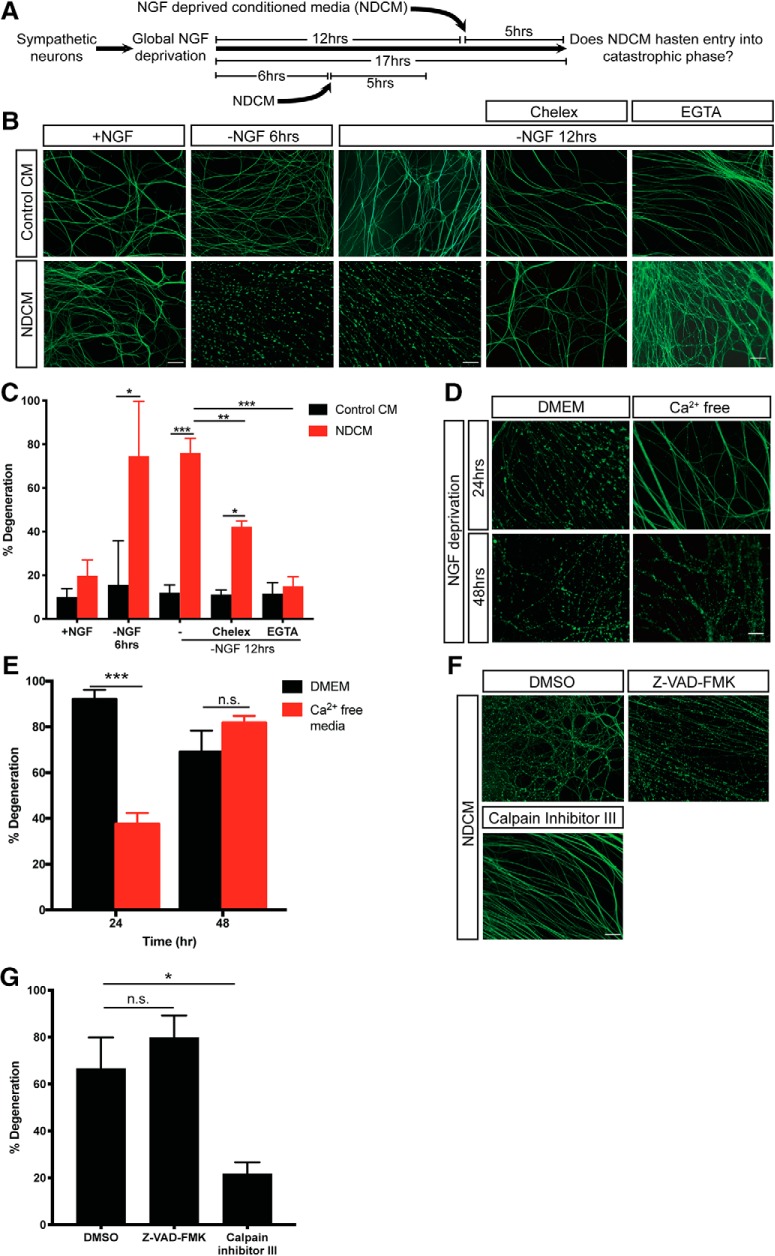
Calcium and calpain activation are required for NDCM-induced catastrophic axon degeneration. A, Conditioned media degeneration paradigm. WT sympathetic neurons were globally deprived of NGF for 6 or 12 h followed by addition of conditioned media to the axons for 5 h. NDCM was collected from degenerated axons after 24 h of NGF deprivation. B, C, Representative images (B) and quantification (C) of β3-tubulin-immunostained distal sympathetic axons after treatment with Control CM and NDCM for 5 h in the presence and absence of NGF, Chelex beads, and EGTA (6 mm). n = 4 or more for each group. For each repeat, at least 100 axons were scored for degeneration. Compared with Control CM, p = 0.0212 for −NGF 6 h, NDCM; p < 0.0001 for −NGF 12 h, NDCM; p = 0.0021 for −NGF 12 h, Chelex, NDCM. Compared with −NGF 12 h, NDCM, p = 0.0002 for −NGF 12 h, Chelex, NDCM; p < 0.0001 for −NGF 12 h, EGTA, NDCM (two-way ANOVA with Sidak's multiple-comparisons test). D, E, Representative images (D) and quantification (E) of β3-tubulin-immunostained distal sympathetic axons after 24 and 48 h of NGF deprivation in media with calcium (DMEM) and calcium-free media. Axons were cultured in regular media (DMEM) and then switched to calcium-free media at the time of NGF deprivation. n = 3 for each group. For each repeat, at least 100 axons were scored for degeneration. p < 0.0001 for 24 h, p = 0.4766 for 48 h (two-way ANOVA with Sidak's multiple-comparisons test). F, G, Representative images (F) and quantification (G) of β3-tubulin-immunostained distal sympathetic axons after treatment with NDCM for 5 h in the presence of DMSO, 50 μm Z-VAD-FMK, and 20 μm calpain inhibitor III, respectively. All cultures were globally deprived of NGF for 12 h before NDCM incubation. Compared with DMSO (n = 7), p = 0.5328, n = 9 for Z-VAD-FMK; p = 0.0080, n = 8 for calpain inhibitor III (one-way ANOVA with Dunnett's multiple-comparisons test). Data are mean ± SEM. *p < 0.05, **p < 0.001, ***p < 0.0001. Scale bar, 50 μm.

