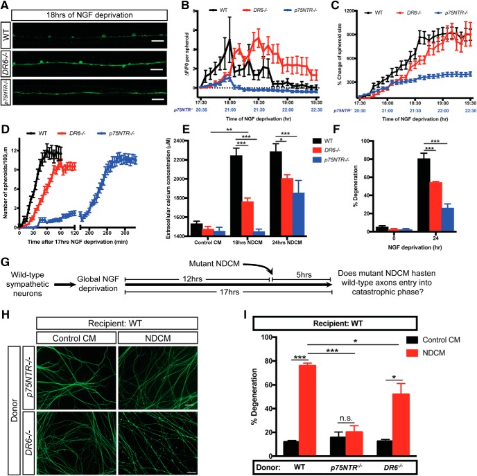
Figure 5.
Depletion of p75NTR delays formation of spheroids and prodegenerative molecule exclusion after NGF deprivation. A, Fluo4-AM calcium imaging of WT, DR6−/−, and p75NTR−/− sympathetic axons 18 h of NGF deprivation. Scale bar, 10 μm. B, C, Spheroidal calcium fluorescence (B) and size change (C) of WT, DR6−/−, and p75NTR−/− sympathetic axons after indicated time of NGF deprivation. B, Horizontal dotted line indicates the baseline without any calcium change. Lower x axis labeled with blue correlates with time of NGF deprivation in p75NTR−/− sympathetic axons. Individual axonal spheroids were quantified: n = 32 (WT), n = 30 (DR6−/−), and n = 22 (p75NTR−/−) spheroids from cultured neurons harvested from three independent litters. D, Quantification of axonal spheroid number per 100 μm of WT, DR6−/−, and p75NTR−/− sympathetic axon at the indicated times after 17 h of NGF deprivation. Individual axonal spheroids were counted: n = 20 (WT), n = 29 ((DR6−/−), and n = 22 (p75NTR−/−) axons from cultured neurons harvested from three independent litters. E, Measurement of extracellular calcium concentration of +NGF (Control CM), 18 and 24 h NDCM collected from WT, DR6−/−, and p75NTR−/− sympathetic axons. n = 4 or more for each group. Compared with WT, 18 h NDCM, p < 0.0001 for DR6−/−, 18 h NDCM; p < 0.0001 for p75NTR−/−, 18 h NDCM. Compared with WT, 24 h NDCM, p = 0.0026 for DR6−/−, 24 h NDCM; p < 0.0001 for p75NTR−/−, 24 h NDCM. Compared with DR6−/−, Control CM, p = 0.0045 for DR6−/−, 18 h NDCM (two-way ANOVA with Dunnett's multiple-comparisons test). F, Quantification of degeneration of WT, DR6−/−, and p75NTR−/− sympathetic axons with or without 24 h of NGF deprivation. n = 3 or more for each group. For each repeat, at least 100 axons were scored for degeneration. Compared with WT, 24 h, p < 0.0001 for DR6−/−, 24 h; p < 0.0001 for p75NTR−/−, 24 h (two-way ANOVA with Dunnett's multiple-comparisons test). G, Catastrophic axon degeneration paradigm to test the prodegenerative effect of mutant NDCM. WT sympathetic neurons were globally deprived of NGF for 12 h followed by addition of conditioned media derived from mutant axons for 5 h. Mutant NDCM was collected from degenerating p75NTR−/− or DR6−/− axons 24 h after NGF deprivation. H, I, Representative images (H) and quantification (I) of WT distal sympathetic axons immunostained for β3-tubulin after treatment with Control CM and NDCM collected from p75NTR−/− and DR6−/− axons for 5 h. Scale bar, 50 μm. Left two columns represent percentages of degeneration of WT sympathetic axons treated with WT NDCM and Control CM, respectively. Compared with WT, NDCM (n = 10), p < 0.0001, n = 7 for p75NTR−/−, NDCM; p = 0.0010, n = 7 for DR6−/−, NDCM (two-way ANOVA with Dunnett's multiple-comparisons test). Compared with Control CM, p < 0.0001 for WT, NDCM; p = 0.8980 for p75NTR−/−, NDCM; p = 0.0014 DR6−/−, NDCM (two-way ANOVA with Sidak's multiple-comparisons test). Data are mean ± SEM. *p < 0.05, **p < 0.001, ***p < 0.0001.

