
Figure 3.
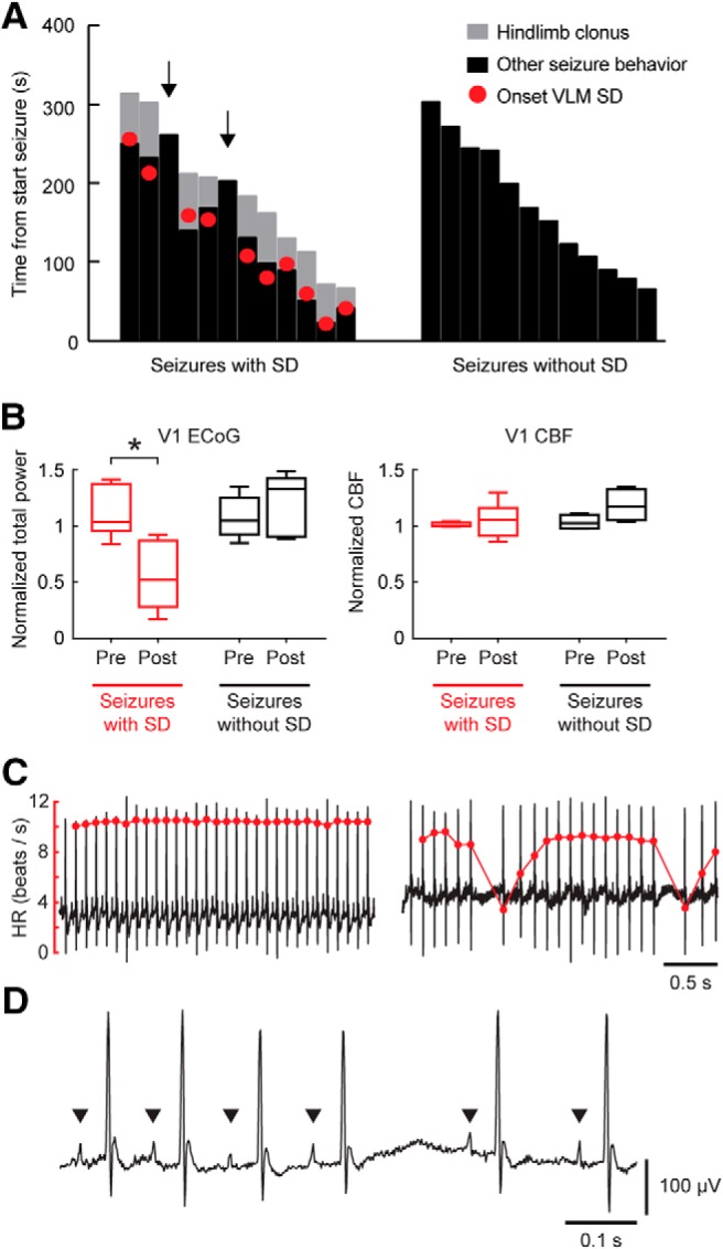
Seizure behavior and post-ictal dynamics indicate brainstem SD during nonfatal seizures in Cacna1aS218L mice. A, Duration of hindlimb clonus and other seizure behavior for seizures with and without brainstem SD, indicated from onset of seizure behavior (at time = 0 s). Hindlimb clonus only occurred during seizures with SD in VLM, and started closely to SD onset. Note that hindlimb clonus did not occur during seizures without brainstem SD and seizures with SD restricted to the PnO (n = 2; arrows). B, ECoG power was significantly suppressed during the post-ictal period for seizures with brainstem SD (t(7) = 5.74, *p = 0.001, paired t test), whereas no difference was present for seizures without brainstem SD. CBF was not significantly different (V1, primary visual cortex). C, Examples of ECG signal (black) and heart rate (HR; red) during a pre-ictal (left) and post-ictal (right) period of a nonfatal seizure with brainstem SD, showing bradyarrhythmias. D, Example of post-ictal ECG signal showing P waves (indicated by arrowheads) and their absence during a prolonged R-R pause.