Figure 1.
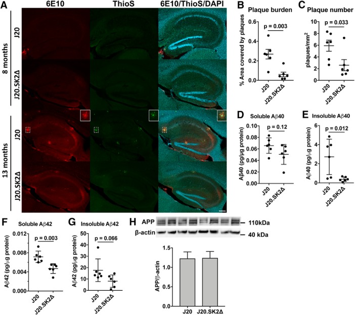
Loss of SK2 greatly reduces Aβ production. A, Immunofluorescence colabeling with 6E10 antibody (red), thioflavin S (ThioS; green), and DAPI (blue) in hippocampus of J20 and J20.SK2Δ mice at 8 and 13 months of age. Box (closed border) shows an enlarged view of an Aβ plaque (dashed box). Scale bar, 200 μm. B, Aβ plaque burden (percentage hippocampal area covered by plaques) and (C) Aβ plaque number in 13-month-old mice. D, E, Aβ40 and (F, G) Aβ42 levels in hippocampus of 13-month-old J20.SK2Δ or J20 mice, as determined by ELISA. Aβ levels were below the limit of detection in WT or SK2Δ mice. H, Western blot for full-length APP in J20 and J20.SK2Δ mice. Statistical significance in B–H was determined by two-tailed, unpaired t tests (6 mice per group).

