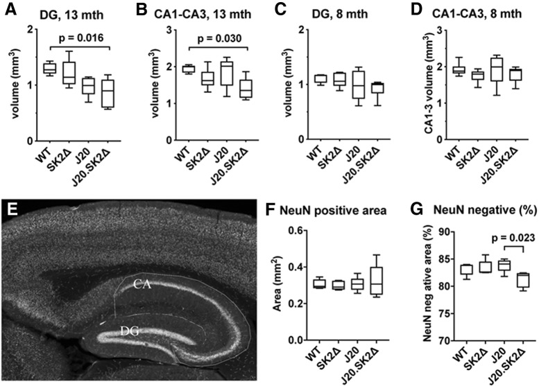
Figure 4.
Loss of hippocampal volume in J20.SK2Δ mice. A–D) Estimated volume of the DG (A, C) and CA1–3 region (B, D) of the hippocampus in 13-month-old (A, B) and 8-month-old (C, D) mice. mth, Month. E, NeuN staining of a WT mouse hippocampus with CA and DG regions outlined. F, Mean NeuN-positive area of the hippocampus in 13-month-old mice. G, NeuN-negative area as a percentage of total hippocampal area in 13-month-old mice. Statistical significance was determined by one-way ANOVA with Sidak's post-test (5–6 mice per group). ANOVA results reported in-text with significant post-test results shown on the graphs.

