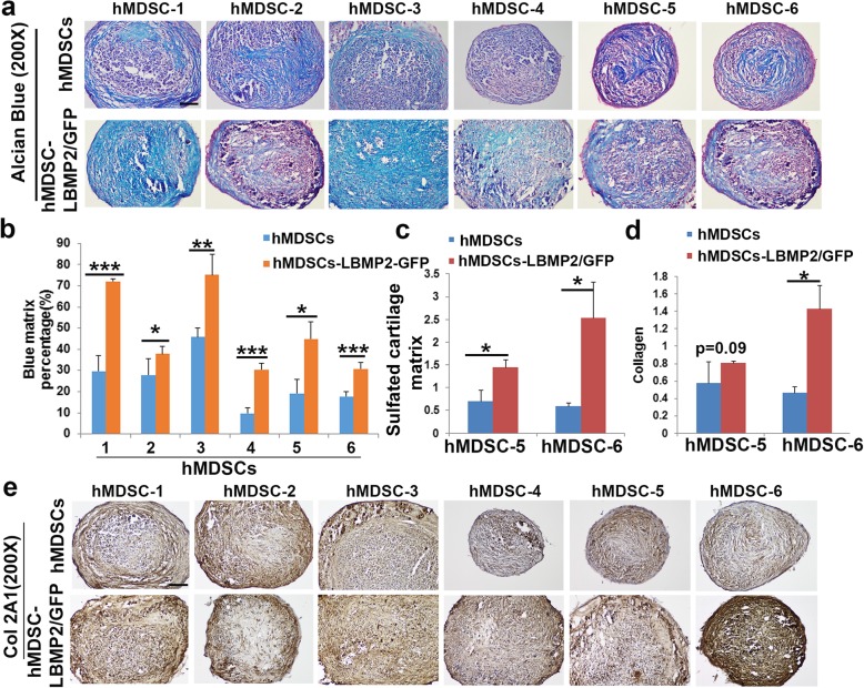
Fig. 2.
a Alcian blue staining. Cartilage matrix including acidic sulfated mucosubstances and hyaluronic acid are stained in blue. b Quantification of blue matrix showed significantly higher percentages of blue matrix in the LBMP2/GFP-transduced cells compared to non-transduced hMDSCs. Scale bar = 100 μm. c Raman spectroscopy quantification indicated significantly higher amount of sulfate cartilage matrix (proteoglycan aggrecan) in LBMP2/GFP-transduced hMDSCs than in non-transduced hMDSCs. d Raman spectroscopy indicated higher collagen content in the LBMP2/GFP-transduced hMDSCs, although differences were not always significant. e Collagen 2A1 (Col2A1) immunohistochemistry indicated stronger Col2A1 staining in the LBMP2/GFP-transduced hMDSCs compared to respective non-transduced hMDSCs. Scale bar = 100 μm. *P < 0.05, **P < 0.01, ***P < 0.001

