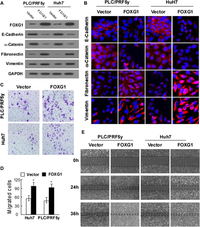
Fig. 2.
Overexpression of FOXG1 promotes cell mobility and invasion by inducing epithelial-mesenchymal transition (EMT). a Western blotting analysis and b Immunofluorescence analysis of expression of epithelial cell markers (E-cadherin and α-catenin) and mesenchymal cell markers (vimentin and fibronectin) in indicated cells transfected with FOXG1 expression vector or control vector. Nuclei counterstained with DAPI. GAPDH was used as a loading control. c Representative migrating images of the indicated Huh7 and PLC/PRF5y cells on uncoated Transwell devices in five random fields. d Quantification of the invading cells of the indicated Huh7 and PLC/PRF5y cells on Matrigel-coated Transwell devices in five random fields. Values represent mean ± SD. *P < 0.05. e Representative micrographs of wound healing assay of the indicated Huh7 and PLC/PRF5y cells. Wound closures were photographed at 0, 24, and 36 h after wounding. All experiments were repeated at least three times with similar results

