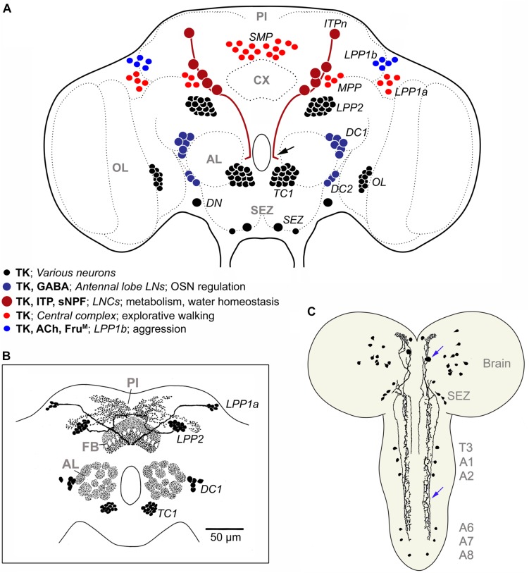
FIGURE 8.
TK in the Drosophila brain. (A) Schematic of neuronal TK distribution in the adult Drosophila brain (frontal view). Neuronal cell bodies are shown in different colors (see legend in figure) to indicate those that have been studied functionally in some detail (blue and red shades), versus those that remain unexplored (black). The light red neurons (SMP, MPP, LPP1a) innervate different layers of the fan-shaped body of the central complex (Kahsai and Winther, 2011) and modulate explorative walking (Kahsai et al., 2010b). The dark blue ones (DC1, DC2) are local neurons of the antennal lobe, some of which coexpress GABA, and are part of circuitry that regulates odor sensitivity in olfactory sensory neurons (OSNs) (Ignell et al., 2009). In male flies the light blue neurons (LPP1b) express FruM and probably acetylcholine (Ach) and regulate levels of aggression (Asahina et al., 2014). The dark red ones (ITPn) are lateral neurosecretory cells (LNCs) with axon terminations in the corpora cardiaca-corpora allata, anterior aorta and intestine (Kahsai et al., 2010a). These cells (ITPn) co-express TK, ion transport peptide (ITP) and short neuropeptide F (sNPF) and regulate aspects of metabolic and water homeostasis (Kahsai et al., 2010a; Galikova et al., 2018). Arrow indicates axon destined for retrocerebral complex of the black neurons LPP2 and TC1 neurons send axons to the pars intercerebralis (PI) and dorsal protocerebrum (Lundquist et al., 1994; Winther et al., 2003), the DNs were assumed to be descending neurons (Winther et al., 2003), and resemble natalisin-producing ICLI neurons shown in Supplementary Figure S5 (Jiang et al., 2013). The branching of the neurons associated with the optic lobes (OL) and subesophageal zone (SEZ) has not been unraveled. The terminology (except ITPn) is from Winther et al. (2003) and specification of neurons compiled from papers cited above. (B) Schematic of TK distribution in some neuropil regions of the Drosophila brain. FB, fan-shaped body, other acronyms as in (A). Modified from Nässel (2002). (C) TK immunoreactive neurons in brain and ventral nerve cord of third instar larva of Drosophila, slightly edited from Winther et al. (2003). Blue arrows indicate a descending neuron. T3, third thoracic neuromere; A1-A8 abdominal neuromeres.

