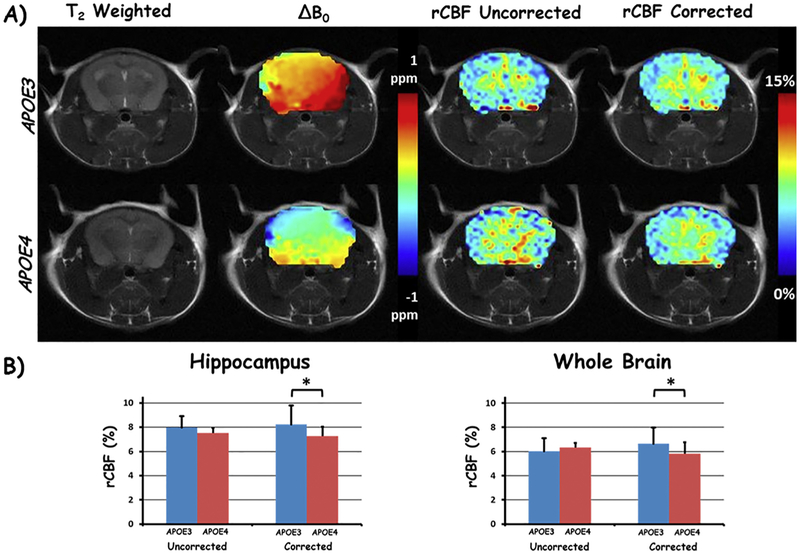
Fig. 6.
TADDZ correction produced B0-corrected CBF maps can differentiate the subtle CBF difference between APOE3 and APOE4 genotyped Alzheimer's disease. A) Representative brain images from an APOE3, top row, and an APOE4, bottom row, mouse. Columns from left to right are T2 weighted image, ΔB0, uncorrected (from conventional ASL MRI), and corrected rCBF maps with TADDZ, respectively. B) Bar chart depiction of the rCBF differences between APOE3 and APOE4 mice, uncorrected (conventional ASL MRI, left) and corrected (TADDZ MRI, right) within the hippocampus (left chart) and whole brain (right chart). *p < 0.05.

