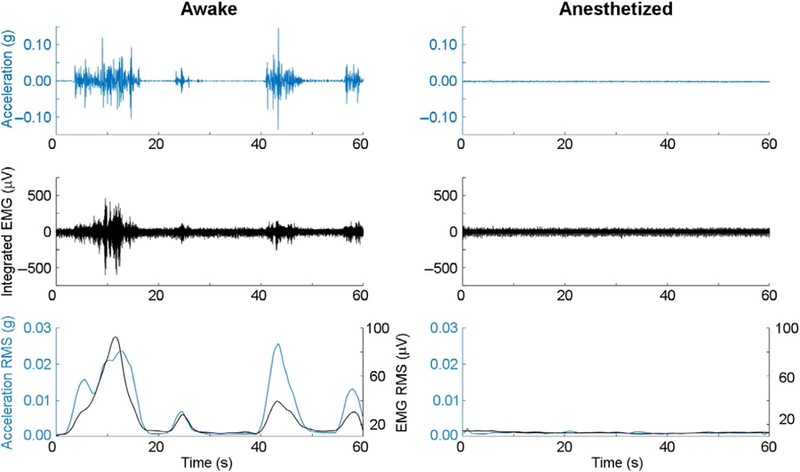
Fig. 5.

Analysis of acceleration and EMG are useful modalities to determine duration of anesthetic sedation. One-minute raw acceleration (Row 1) and electromyographic traces (Row 2) are shown before (Left) and after (Right) a 1.2% isoflurane anesthetic exposure for an individual mouse. RMS moving average filtering of the raw data shows high correlation (>0.7) between accelometry and electromyography for movement detection (Row 3).
