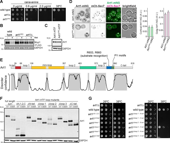
FIGURE 1:
Art1 contains an arrestin domain disrupted by multiple insertions. (A) Serial dilutions of the indicated yeast strains spotted on synthetic media containing canavanine. (B) Immunoblot of the yeast strains in A expressing Mup1-FLAG before and after treatment with 20 µg/ml Met for 60 min. (C) Immunoblot of yeast expressing Art1 or Art12RD with a C-terminal HTF (6xHis-TEV-3xFLAG) tag. Art1-HTF was detected with an α-FLAG antibody. (D) Localization of Art1-mNG and Art12RD-mNG in minimal media. Scale bar = 2 µm. Right, quantification of Art1 localization at the PM and Golgi; error bars are 95% CI, n > 300 cells for each condition. (E) Top, Art1 schematic. Nonconserved loop regions shown in gray. Conserved regions predicted to form an arrestin fold are colored. Bottom, disorder confidence predicted DISOPRED3. Gray shading indicates predicted disordered regions. (F) Immunoblot of Art1-HTF tail and loop mutants, with and without the K486R mutation, detected with an α-FLAG antibody. (G) Serial dilutions of Art1 tail and loop mutants spotted on synthetic media.

