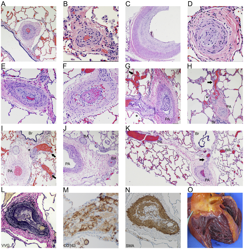
Figure 2: Pathology features consistent with pulmonary hypertension.
H+E stained sections of both small and large arteries show marked muscular hypertrophy (A, B), marked cellular intimal proliferation (C) leading to complete luminal occlusion (D). Also noted was concentric intimal fibrosis (E) and intraluminal thrombus formation (F). H+E sections showed histologic features consistent with severe pulmonary hypertension including plexiform (*) and dilation (arrow) lesions associated with markedly remodeled pulmonary arteries (PA) (G, H, I). Open intrapulmonary bronchopulmonary anastomoses (K) that bridge the pulmonary arteries and bronchial arteries (BA), along with marked BA remodeling and occlusions (J) are present (Br: bronchiole). Microscopic examination of the lungs included elastin [Verhoeff-van Gieson (VVG), L] stained sections showing marked pulmonary artery remodeling. Immunohistochemistry on lung sections revealed fibrointimal proliferation with infiltration of macrophages (CD163+, M) and tunica medical thickening [smooth muscle actin (SMA)+, N]. Gross examination of the heart showing dilatation of the right ventricle (O).

