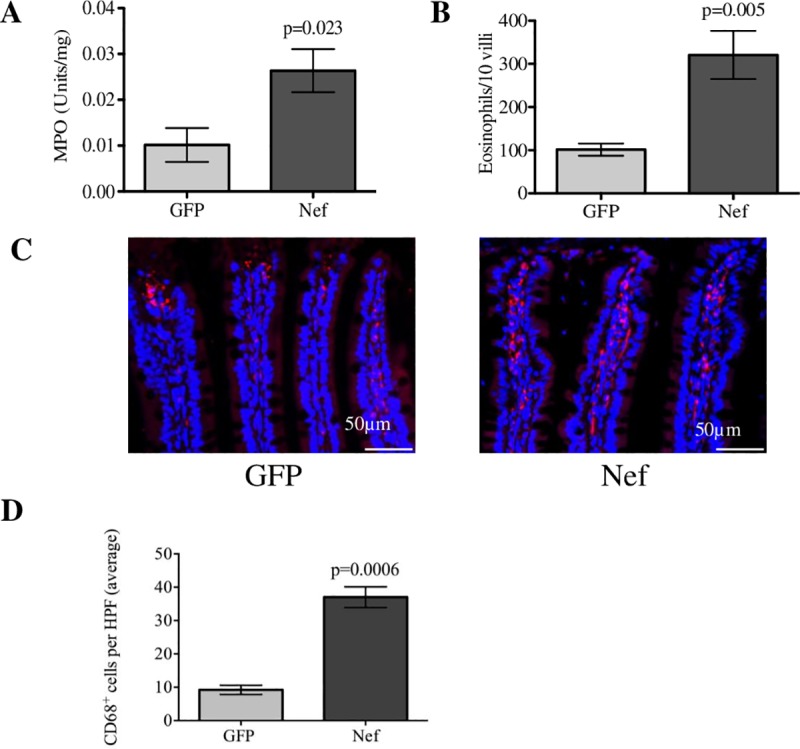
Fig 4. Hippocampal HIV-1 Nef expression induces intestinal inflammation.

(A) An MPO activity assay was employed to assess myeloid infiltration into the ileal area. Nef-treated rats showed increased MPO levels in the ileum compared to the GFP-treated rats. (B) Eosinophils counted from H&E-stained ileal tissue (10 villi per rat) showed that infiltration was significantly increased in the HIV-1 Nef-treated rats. (C) Immunofluorescence for the macrophage marker CD68 (red) was assessed to determine the levels of macrophage infiltration between the groups while nuclei of the cell are represented (in blue). (D) Macrophages were quantified in three 40x HPFs per rat. N = 3 to 7 rats per group. Student’s t-test was performed to determine statistical difference between the Nef-treated group and the GFP-treated group (control). Scale bars indicate 50 μm. Error bars indicate means ± S.E.M.
