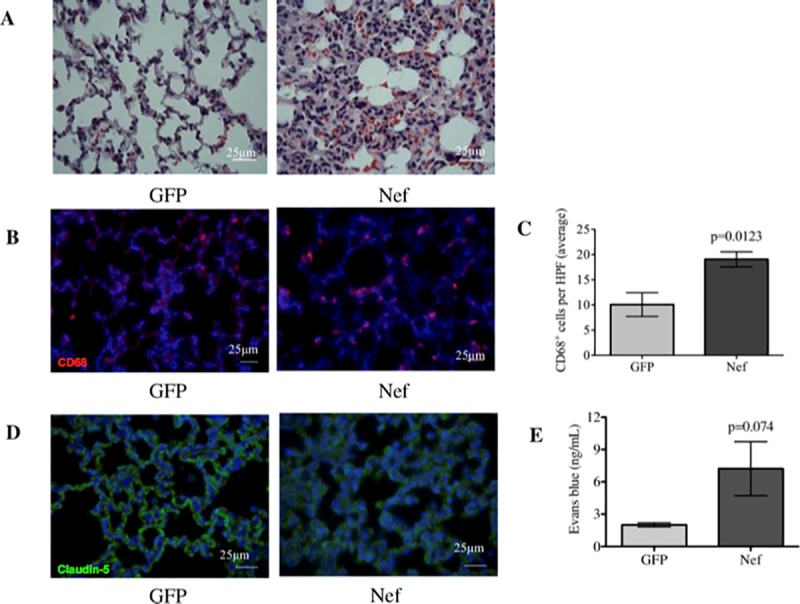
Fig 6. Nef expression in the rat hippocampus induces interstitial pneumonitis and increases macrophage infiltration and vascular permeability in the lung.

(A) H&E staining showed that the lungs from GFP-treated rats appear normal, while the lungs from Nef-treated rats showed marked infiltration of immune cell and interstitial thickening. (B) Immunofluorescence for the macrophage marker CD68 in (red) while nuclei of the cells is represented in (blue) was assessed to compare macrophage infiltration between the groups. (C) Macrophages were quantified in three HPFs per rat. (D) Immunofluorescence of the tight junction protein claudin-5 in (green) while nuclei of the cells is represented in (blue) was assessed to determine tight junction alterations in the lungs. Nef-treated rats showed alterations in claudin-5 expression that were characterized by diffuse and dimmed staining compared to what was observed in the GFP-treated rats. (E) Changes in lung permeability were determined by measuring EB extravasation, which confirmed that Nef-treated rats had increased lung permeability compared to our control group. N = 4 to 6 rats per group. Scale bars indicate 25 μm. Student’s t-test was performed to determine differences between the Nef-treated group and the GFP-treated group (control). Data are expressed as means ± S.E.M.
