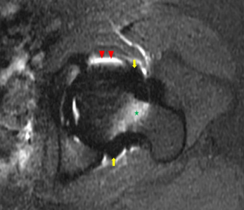
Figure 1. Axial T1 weighted fat-saturated magnetic resonance image of the left hip.
A T1 weighted axial fat-saturated magnetic resonance image of the left hip exhibits a signal void surrounding the left hip (yellow arrows), conforming to the shape of the joint capsule, status post-injection for magnetic resonance arthrography. There is an incidental note of altered fat saturation at the anterior and posterior joint capsule margins (red arrowheads) and within the marrow of the femoral neck (green star).

