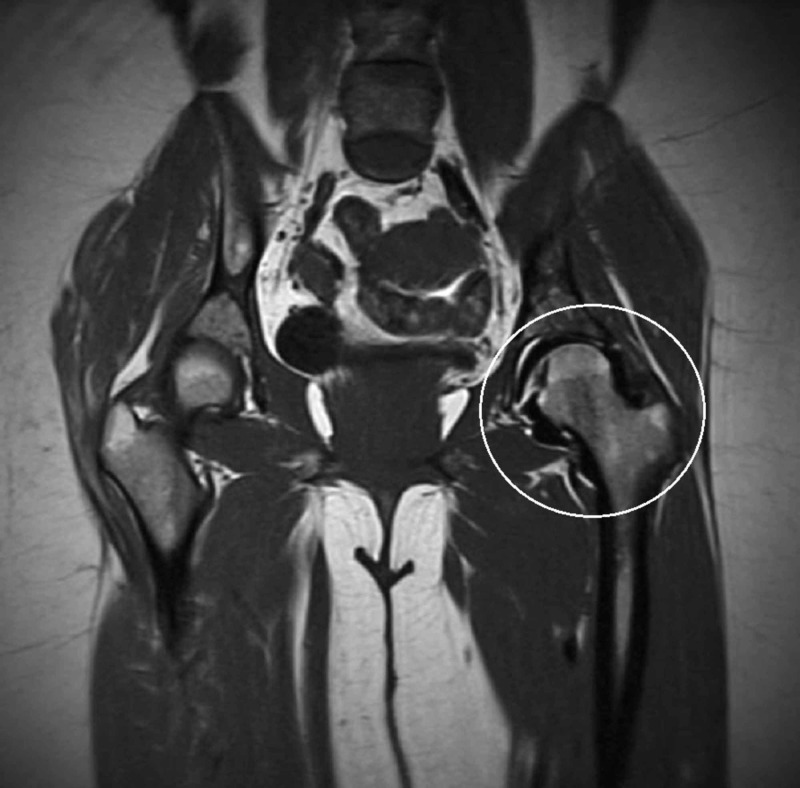
Figure 2. T1 weighted coronal magnetic resonance image of the pelvis.
The T1 weighted coronal magnetic resonance image of the left hip (white circle) exhibits a signal void surrounding the left hip and conforms to the shape of the joint capsule status post-injection for magnetic resonance arthrography.

