Figure 2.
Molecular Cloning and Confirmation of the WP1 Gene.
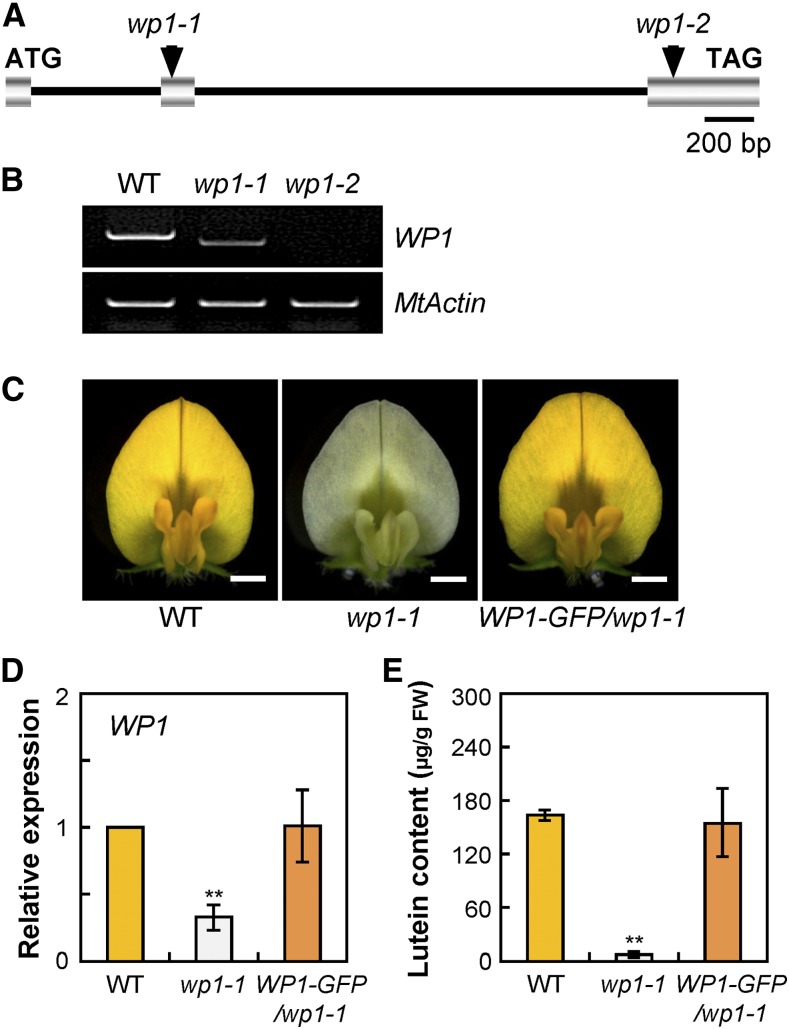
(A) Schematic representation of the gene structure of WP1 showing the Tnt1 insertion sites in wp1-1 and wp1-2. Introns are represented by a line, and exons are represented by a striped box.
(B) RT-PCR showing transcript abundance of WP1 in the flowers of the wild type and wp1 mutants. MtActin was used as the control. WT, wild type.
(C) Genetic complementation of wp1-1. Representative flowers of the wild type, wp1-1, and wp1-1 complemented with WP1-GFP (WP1-GFP/wp1-1). Bars = 1.5 mm. WT, wild type.
(D) Transcript levels of WP1 in petals of the wild type, wp1-1, and WP1-GFP/wp1-1. Bars represent means ± sd of three biological replicates. Asterisks indicate differences from the wild type (**P < 0.01, Student’s t test). WT, wild type.
(E) Lutein concentrations in petals of the wild type, wp1-1, and WP1-GFP/wp1-1. Bars represent means ± sd of three biological replicates. Asterisks indicate differences from the wild type (**P < 0.01, Student’s t test). WT, wild type.

