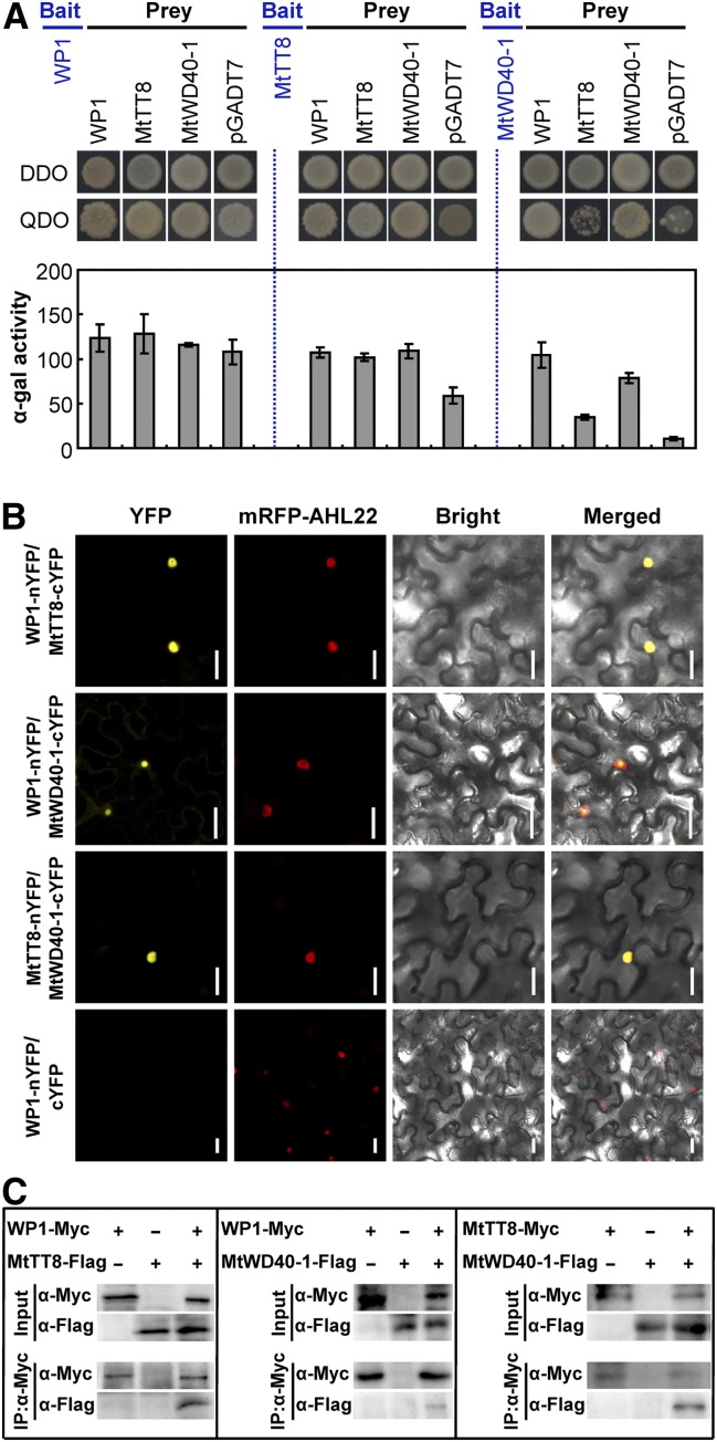
Figure 6.
WP1 Interacts with MtTT8 and MtWD40-1 to Form an MBW Complex.
(A) Interaction between WP1, MtTT8, and MtWD40-1 in the Y2H assay. Plate auxotroph and α-galactosidase assay showing interaction of each protein. Bars represent means ± sd of three independent experiments. α-gal, α-galactosidase; DDO, double dropout; pGADT7, prey plasmid; QDO, quadruple dropout.
(B) Interaction between WP1, MtTT8, and MtWD40-1 in N. benthamiana leaf epidermal cells using a split YFP BiFC assay. AHL22 was used as a nuclear localization marker. Bars = 25 μm.
(C) Interaction between WP1, MtTT8, and MtWD40-1 in N. benthamiana using a Co-IP assay. Immunoblots of the total protein extracts (Input) and the IP product were performed using the anti-Myc antibody (α-Myc) or anti-Flag antibody (α-Flag), respectively.

