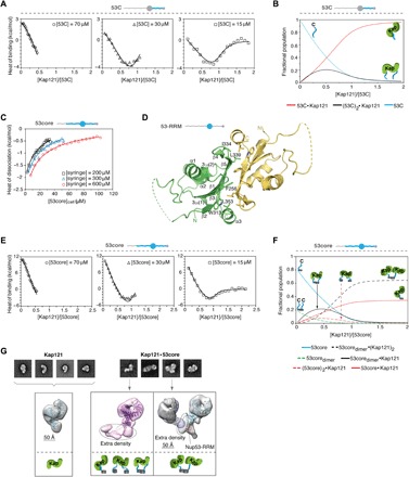
Fig. 2. Nup53 RRM and IDR domains are coupled when interacting with Kaps.

(A) ITC profiles for 53C-Kap121 interactions at three different 53C concentrations. The isotherms were globally analyzed together with the reverse titration in fig. S2D by a two-mode binding model (continuous lines; best-fit parameters with SDs are reported in Table 1). (B) Population distribution of 1:1 and 2:1 53C·Kap121 complexes is displayed as a function of [Kap121]/[53C] molar ratio. (C) 53core monomer-dimer equilibrium at different concentrations analyzed by ITC using a single-step dissociation model (continuous lines). (D) Crystal structure of the yeast Nup53 RRM homodimer determined at a resolution of 1.75 Å. Individual monomers are shown with secondary structure elements. Key hydrophobic residues contributing to the interface are highlighted in the green monomer. Regions not observed in the electron density map are shown as dashed lines. (E) ITC profiles for 53core-Kap121 interactions at three 53core concentrations were globally analyzed by a multiple-equilibrium model (continuous lines; best-fit parameters with SDs are reported in Table 1). (F) Population distribution of the 53core·Kap121 complex is shown as a function of [Kap121]/[53core] molar ratio. (G) Negative-stain EM (NS-EM) analysis of Kap121 and its complex with 53core. Representative 2D class averages and 3D reconstitution of free Kap121 and the two distinct conformations of the 53core·Kap121 complex are displayed with fitted crystal structures of Kap121 (PDB code: 3W3T). The monomeric state of the 53core·Kap121 complex (middle panel) shows extra density that likely represents either a 53core monomer, dimer, or an average of the two conformations, as determined by ITC. To assign orientation of the two Kap molecules within the dimeric state of the complex (right panel), the experimental NS-EM density map for free Kap121 (purple and cyan) was fitted into each of the arms of the structure. The RRM domain of Nup53 (PDB code: 5UAZ) was docked into the stemlike portion of the structure.
