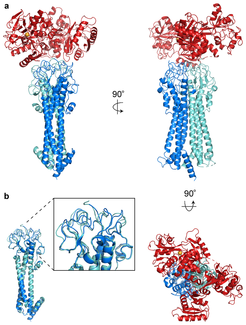
Figure 1. The structure of the trypanosome transferrin receptor.
a. The structure of the trypanosome transferrin receptor heterodimer (ESAG6 in dark blue and ESAG7 in light blue) bound to human transferrin (red). The iron ion is shown as an orange sphere. b. An alignment of ESAG and ESAG7 showing the divergence of the membrane-distal loops to create an asymmetric binding site for transferrin.

