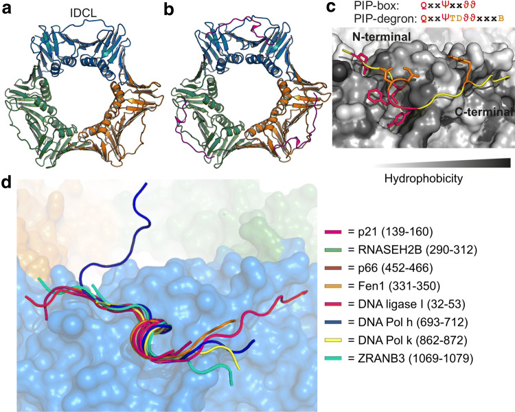
Fig. 1.
PCNA is a circular trimer that binds disordered ligands through short linear motifs. a Structure of unbound human PCNA (PDB-code:1VYM) highlighting the three different subunits colored in blue, orange and green, respectively, and the interdomain connecting loop (IDCL) indicated. b Structure of p21 (magenta) bound to PCNA (PDB-code: 1AXC; coloring as in a). c Magnification of the binding pockets of PCNA with bound p21. The binding pocket is made from residues 40–44, 117–135 (IDCL), 230–235, and 251–253. The PCNA surface is colored in gray shades according to hydrophobicity; the PIP-box residues inserted into the binding pockets are highlighted in red and degron specific residues in orange. d Overlay of seven peptides crystallized in complex with human PCNA including a degron (p21) and an APIM (ZRANB3). The PCNA surface is from the p21-complex (PDB-code: 1AXC) and colored as described in a

