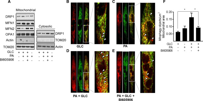
Fig. 8.
Effects of cellular fuel overloading on mitophagy and upon proteins linked to mitochondrial dynamics. Wild type L6 myotubes or those stably expressing a retroviral vector encoding a GFP–mCherry–Fis1 mitophagy reporter were incubated with GLC (5 mM), PA (0.4 mM) and BI605906 (10 μM) for 16 h as indicated prior to: a subcellular fractionation and isolation of a cytosolic and mitochondrial-enriched membrane fraction for immunoblotting with antibodies to proteins shown. b–e Fixing and confocal imaging to visualise and f quantifying (using the Volocity software) mitophagy in L6 myotubes. Data (mean ± SEM) in (f) are from five separate experiments. Asterisks indicate a significant change (P < 0.05) between the indicated bars. Boxed sections in panels b, c, d and e have been expanded to highlight mitophagic particles, some of which are depicted by the white arrow heads

