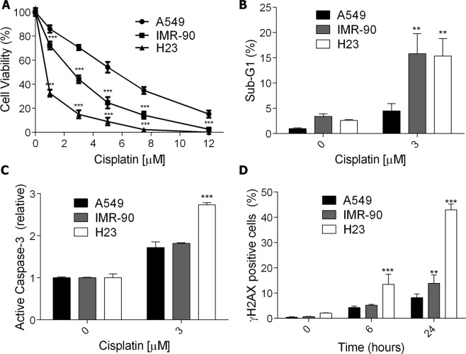
Figure 1.
Cell death and DNA damage induction in normal and cancer lung cells after exposure to cisplatin. (A) A dose-response curve of three lung cell lines treated with increasing concentrations of cisplatin and analyzed after 72 h using the XTT assay. (B,C) The apoptotic fraction of lung cells treated with cisplatin for 72 h, analyzed as the sub-G1 population levels using flow cytometry of PI-stained nuclei or the fold increase of cells with active caspase-3 relative to control. (D) Flow cytometry analysis of γH2AX positive staining in lung cells upon treatment with cisplatin (5 μM) for 6 h and 24 h. Values are mean ± SEM of three independent experiments, *P < 0.05, **P < 0.01, ***P < 0.001.

