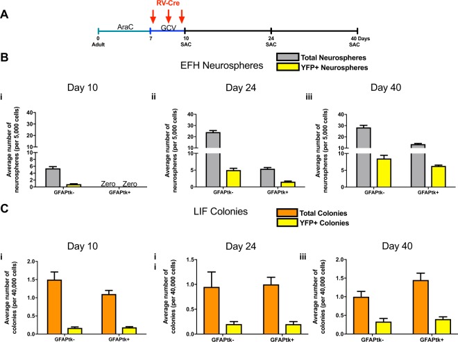
Figure 3.
Proliferating, GFAP negative cells give rise to dNSCs following ablation. (a) Schematic of the experimental paradigm. (b) The neurosphere assay for dNSCs (EFH) (grey bars) performed in control (GFAPtk-) and experimental groups (GFAPtk+) at day 10 (i), day 24 (ii), or day 40 (iii) after onset of ablation. The numbers of YFP+ neurospheres are indicated in yellow bars (n = 6 mice/group/survival time). (c) Colony-forming assay for pNSCs (LIF) (orange bars) performed in control (GFAPtk−) and experimental groups (GFAPtk+) at day 10 (i), day 24 (ii), or day 40 (iii) after the onset of ablation. The numbers of YFP+ colonies are indicated in yellow (n = 6 mice/group for day 10; n = 3 mice/group for day 24; n = 4–6 mice/group for day 40). All data represent mean ± SEM.

