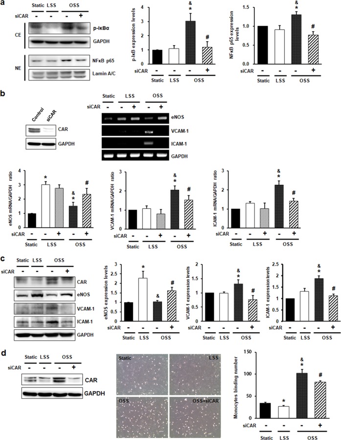
Fig. 3. CAR knockdown inhibits disturbed flow-induced inflammatory responses in endothelial cells.
HUVECs transfected with siRNA against CAR were exposed to FSS for 6 h (b) or 24 h (a). a Cytosolic and nuclear fractions were separated after 24 h of FSS. The extent of IκB phosphorylation and nuclear translocation of NF-κB were determined by western blotting with antibodies against p-IκB, NF-κB p65, GAPDH, and Lamin A/C. GAPDH and Lamin A/C were used as loading controls for the cytosolic and nuclear fractions, respectively. Representative images are shown (n = 5; *P < 0.05, static vs. LSS or OSS; &P < 0.05, LSS vs. OSS; #P < 0.05, OSS vs. siCAR + OSS). b The knockdown of CAR by siCAR was validated by western blotting. Levels of mRNAs encoding anti-inflammatory genes (eNOS), proinflammatory genes (VCAM-1 and ICAM-1), and GAPDH (internal control) were measured by RT-PCR. Representative images are shown (n = 5; *P < 0.05, static vs. LSS or OSS; #P < 0.05, OSS vs. siCAR + OSS). c The protein levels of eNOS, VCAM-1, and ICAM-1 were measured by western blotting. Representative images are shown (n = 5; *P < 0.05, static vs. LSS or OSS; &P < 0.05, LSS vs. OSS; #P < 0.05, OSS vs. siCAR + OSS). d To assay monocyte binding to endothelial cells, we exposed transfected HUVECs to FSS for 24 h. Monocytes in five random optical fields per sample were counted (n = 5; *P < 0.05, static vs. LSS or OSS; &P < 0.05, LSS vs. OSS; #P < 0.05, OSS vs. siCAR + OSS).

