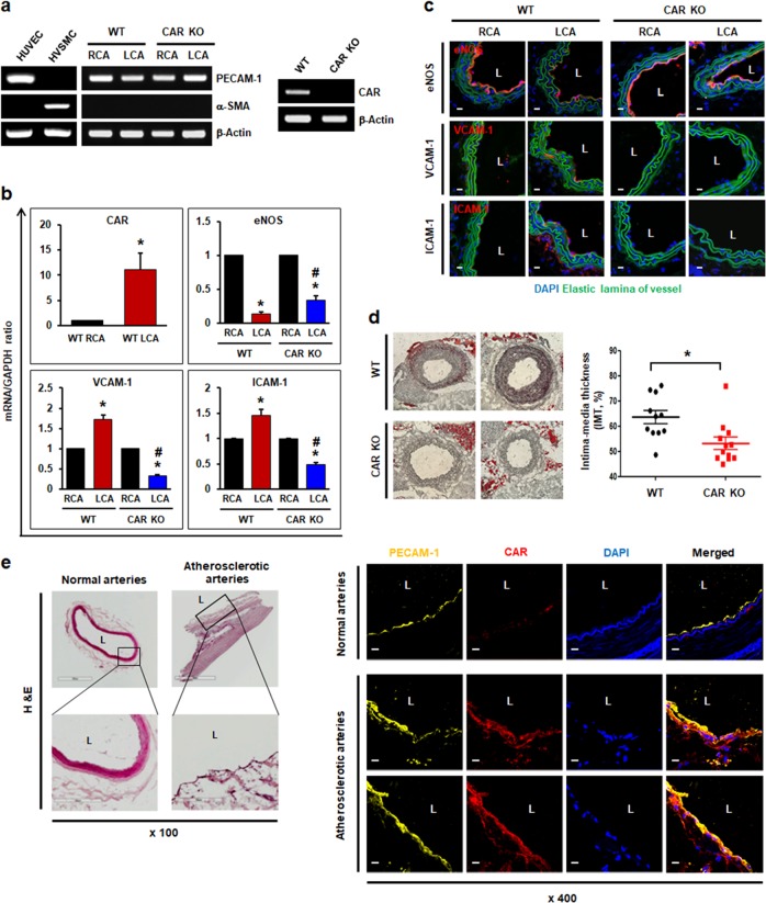
Fig. 4. CAR deletion protects the proatherogenic endothelium under disturbed flow.
Wild-type (WT) C57BL/6 or EC-specific CAR KO mice were partially ligated in the LCA. a Twenty-four hours after LCA ligation, the mice were euthanized, and EC-enriched RNA was extracted from both carotid arteries. EC-enriched RNA was confirmed by assessing an endothelial-specific marker (PECAM-1), a smooth muscle cell-specific marker (α-smooth muscle actin [α-SMA]), and β-actin (internal control). HUVECs were used as a positive control for PECAM-1 and a negative control for α-SMA. Human vascular smooth muscle cells (VSMCs) were used as a positive control for α-SMA and a negative control for PECAM-1. The knockout of endothelial CAR in EC-specific CAR KO mice was confirmed by RT-PCR. Representative images are shown (n = 5). b Levels of mRNAs encoding eNOS, VCAM-1, and ICAM-1 in EC-enriched RNA were determined by real-time PCR (n = 5; *P < 0.05, WT RCA vs. WT LCA, CAR KO RCA vs. CAR KO LCA; #P < 0.05, WT LCA vs. CAR KO LCA). c Immunofluorescence staining was performed in carotid arteries from WT or EC-specific CAR KO mice. Representative images are shown (n = 3; red, eNOS, VCAM-1, or ICAM-1; blue, nuclei; green, elastic lamina of vessel; scale bars, 10 μm). d Disturbed flow-induced atherosclerotic plaque formation was visualized in ligated LCA by Oil Red O staining. We measured the IMT by calculating the luminal area relative to the total vascular area. Representative images are shown (n = 11; *P < 0.05, WT vs. CAR KO). e Human arterial tissues were stained with hematoxylin and eosin (H&E) and anti-PECAM-1 and anti-CAR antibodies. PECAM-1 was used as an endothelial-specific marker. Expression of CAR was compared between normal and atherosclerotic regions of arterial tissue by confocal microscopy. Representative images from at least three experiments are shown (red, CAR; yellow, PECAM-1; blue, nuclei). Scale bars, 10 μm.

