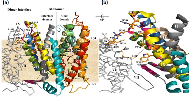
Figure 1.
The NhaA dimer interface. (a) The crystal structure of the NhaA monomer and the interfacial domain between the two monomers of the NhaA dimer are represented according to17 (PDB.ID: 4ATV); one monomer is shown in colored ribbon, and the trans membrane segment (TM) are numbered by white Roman numerals. The relevant residues are in stick representation. Part of the other monomer of the dimer is shown in a grey line; the TMs are numbered in black, and the relevant residues are shown in line representation. In the NhaA dimer interface, sites where single Cys replacements on TM IX and the β-hairpin loop cross-link22,26 are marked in dark pink. The membrane is depicted in wheat color. The cytoplasmic funnel (dots) is composed of TMSs II, IVc (c and p denote cytoplasmic and periplasmic sides, respectively), V, and IX. The periplasmic funnel (dots) comprises TMs II, VIII and XIp. The TM IV/XI assembly of short helices connected by extended chains is shown together with the putative Na+ binding site (D163, D164). Cyt, cytoplasm; Per, periplasm. (b) The direct contacts between NhaA monomers17: W258 and V254 on TM IX of one monomer make a cross interface bridge to TM VII of the other monomer at R204. The two representations were generated using PyMOL with equal notifications.

