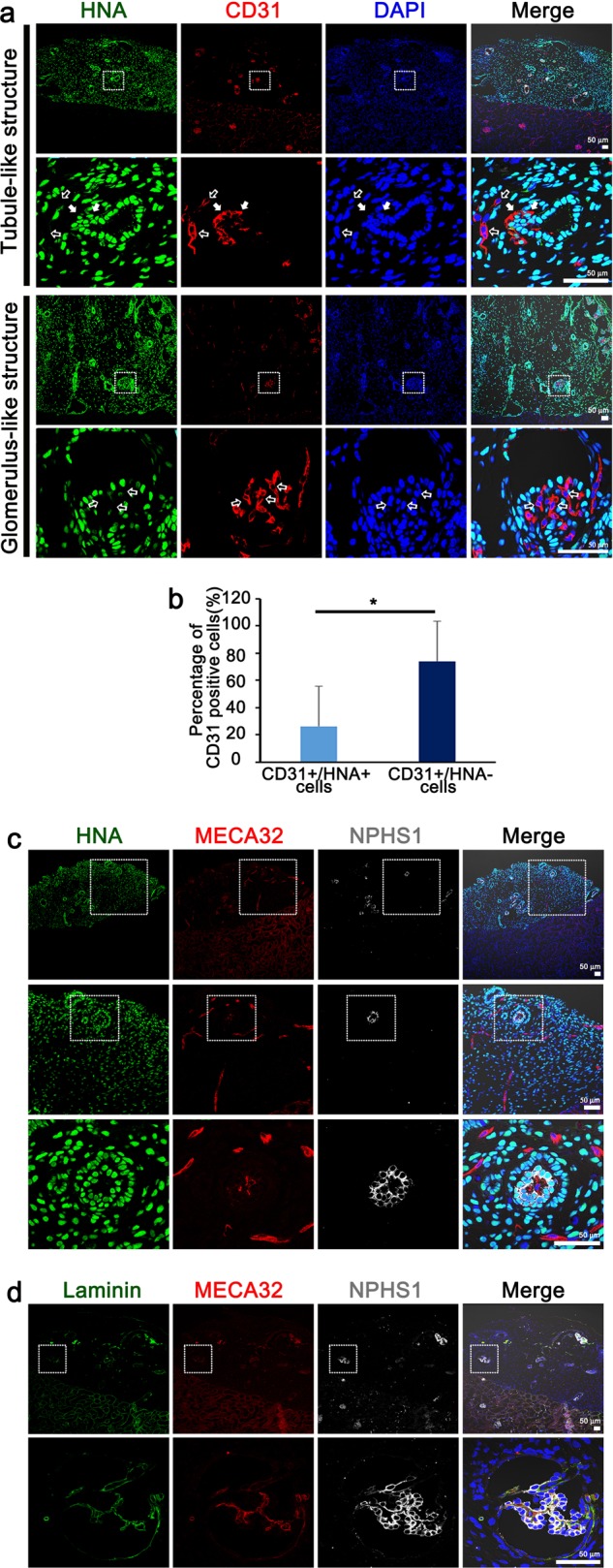
Fig. 3. Transplanted kidney organoids are vascularized with infiltrating endothelial cells from the host kidney and form GBM-like structures.

a Representative confocal images of transplanted kidney organoids at 14 days after transplantation. White arrow in the enlarged images indicate CD-31-positive, HNA-positive cells and open arrow indicate CD-31-positive, HNA-negative cells. Scale bars, 50 μm. b Quantification of CD31+/HNA+ cells and CD31+/HNA− cells of the CD31+ cells. c Representative confocal images of podocytes (NPHS1) and mouse endothelial cells (MECA32) in the transplanted kidney organoids at 14 days after transplantation. Scale bars, 50 μm. d Representative confocal images of glomerular-like structures in the transplanted kidney organoids. Cells were stained for NPHS1, MECA32 and laminin. Scale bars, 50 μm.
