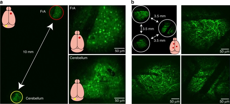
Fig. 3. Demonstration of MATRIEX imaging: structural imaging in multiple brain areas in vivo.
a Left image: a full-frame image including two FOVs in the frontal association cortex (FrA) and the cerebellum. The red and yellow circles indicate two FOVs that are digitally enlarged and shown in the upper-right and lower-right images. A GAD67-GFP transgenic mouse (with the interneurons labeled brain-wide) was used. Two MOs (‘standard version’) were placed at the same depth under a DO (Mitutoyo ×2/0.055). b Example configuration of three FOVs in the cortex of a Thy1-GFP transgenic mouse (with layer 5 cortical neurons specifically labeled and with tuft dendrites visible near the cortical surface). Three MOs (‘standard version’) were placed at the same depth under a DO (Olympus ×4/0.1).

