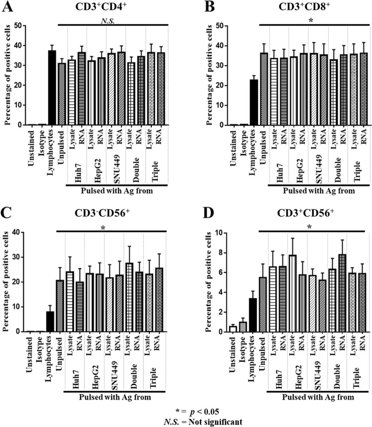
Figure 2.
Expression of lymphocyte markers on effector T-lymphocytes after activation with DCs pulsed with antigens. Autologous lymphocytes (non-adhering PBMCs) were activated to become effector T-lymphocytes by co-culturing with DCs pulsed with cell lysate or RNA prepared from single, double, or triple HCC cell lines for 2 days. Effector T-lymphocytes were then further propagated in AIM-V medium supplemented with IL2, IL7, and IL15 for 10 days. The expressions of CD3+, CD4+, CD8+, and CD56+ were examined by flow cytometry after staining with specified antibodies. The percentages (mean ± SEM) of specific populations of effector cells, including CD3+ CD4+ helper T-cells (A), CD3+ CD8+ cytotoxic T-cells (B), CD3-CD56+ NK cells (C), and CD3+ CD56+ NKT cells (D) were calculated from three independent experiments. (*p < 0.05 compared with non-adhering PBMCs. Data analyzed by one-way ANOVA with Tukey’s correction).

