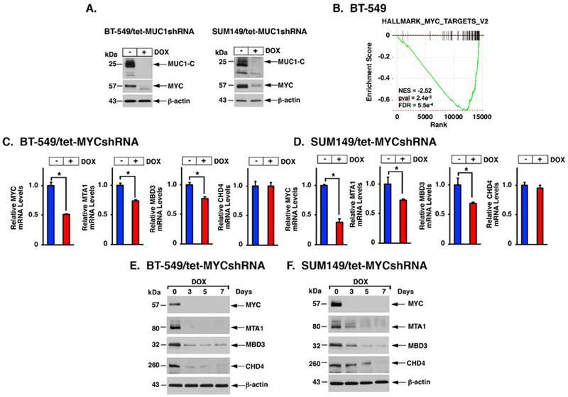
Figure 2. MUC1-C induces MTA1, MBD3 and CHD4 by a MYC-mediated pathway.

A. BT-549/tet-MUC1shRNA (left) and SUM149/tet-MUC1shRNA (right) cells were left untreated or treated with 500 ng/ml DOX for 7 days. Lysates were immunoblotted with antibodies against the indicated proteins. B. RNA-seq was performed in triplicate on BT-549/CshRNA and BT-549/MUC1shRNA cells. The datasets were analyzed with GSEA, using the Hallmark gene signature collection. Silencing MUC1 was significantly associated with suppression of MYC target gene expression. C-D. BT-549 (C) and SUM149 (D) cells expressing a tet-MYCshRNA were left untreated or treated with 500 ng/ml DOX for 7 days. Cells were analyzed for MYC, MTA1, MBD3, CHD4 and HDAC1 mRNA levels. The results (mean±SD) are expressed as relative mRNA levels compared to that obtained for untreated cells (assigned a value of 1). E-F. BT-549/tet-MYCshRNA (E) and SUM149/tet-MYCshRNA (F) cells were treated with 500 ng/ml DOX for the indicated days. Lysates were immunoblotted with antibodies against the indicated proteins.
