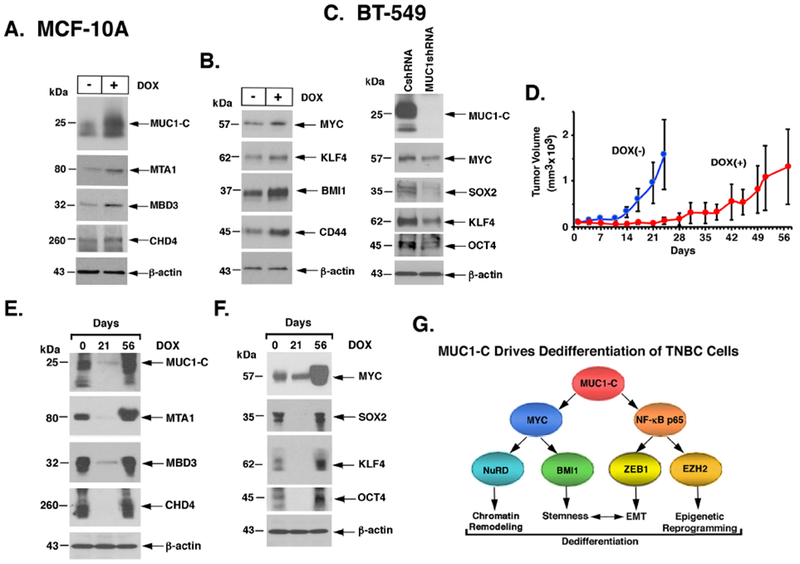
Figure 7. MUC1-C drives dedifferentiation of TNBC cells.

A and B. MCF-10A cells expressing tet-MUC1-C (MCF-lOA/tet-MUC1-C) were left untreated or treated with 500 ng/ml DOX for 7 days. Lysates were immunoblotted with antibodies against the indicated proteins. C. Lysates from BT-549 cells expressing a CshRNA or MUC1shRNA#2 were immunoblotted with antibodies against the indicated proteins. D-F. Female nude mice were injected subcutaneously with SUM149/tet-MUC1shRNA cells. Mice pair-matched into two groups when tumor volumes reached 100 mm3 were fed without (blue) and with DOX (red) for 56 days when the study was terminated. Tumor volumes are expressed as the mean±SD for 6 mice (D). Lysates from tumors harvested on days 0, 21 and 56 were immunoblotted with antibodies against the indicated proteins (E and F). G. Proposed model depicting the role of MUC1-C in integrating NuRD activation with dedifferentiation and plasticity of TNBC cells. MUC1-C has been linked to activation of the IKK→NF-κB p65 and MYC pathways. The disordered MUC1-C cytoplasmic domain binds directly to NF-κB p65 and thereby induces expression of (i) ZEB1 with the induction of EMT and (ii) EZH2 with induction of H3K27 methylation. The present studies further demonstrate that the MUC1-C cytoplasmic domain binds to the MYC HLH-LZ domain, which contributes to regulation of the MYC transactivation function. MUC1-C→MYC signaling induces expression of the BMI1 stem cell factor and, as shown in the present work, the NuRD components MTA1, MBD3 and CHD4. In turn, MUC1-C associates with the NuRD complex and integrates EMT, stemness and epigenetic reprogramming with chromatin remodeling in the dedifferentiation of TNBC cells.
