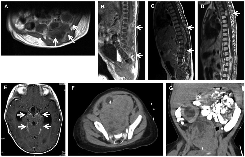
Figure 1.
Radiological findings, with arrow indicating tumor. Gadolinium-enhanced magnetic resonance imaging at first diagnosis (A, B), the first relapse (C), and the second relapse after radiotherapy (D, E) in Patient 1. Patient 2 axial (F) and coronal (G) views from pre-operative computed axial tomography imaging with oral and intravenous contrast showing a heterogenous, paramedian pelvic mass and asymmetric kidneys. There is delayed urinary excretion on the right due to obstruction from the mass. In the axial image (F), the balloon of the urinary catheter is apparent and displaced ventrally and significantly away from the midline.

