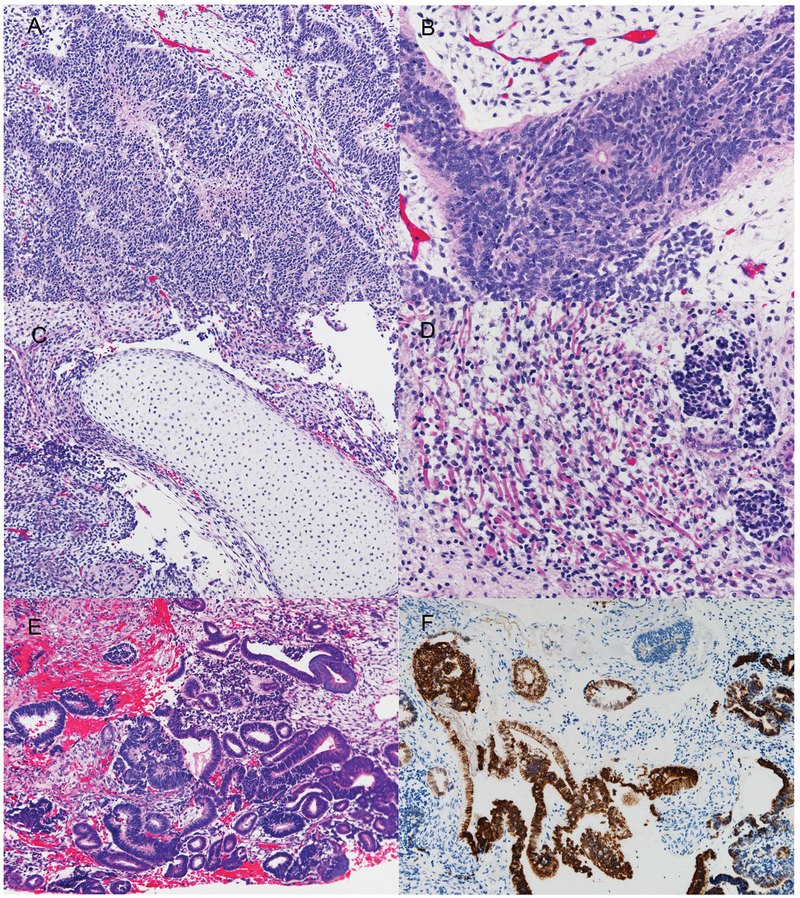
Figure 2.
Microscopic appearance of the multipatterned tumor from Patient 1. The primary tumor from Patient 1 showed immature neuroepithelial components including multilayered rosettes and medulloepithelioma-like component (A, B), and immature mesenchymal tissues, including mature cartilage and skeletal muscle cells (C, D). The recurrent tumor was composed predominantly of neuroepithelial tubular structures in loose mesenchymal tissue (E). These tubules were positive for cytokeratin CAM 5.2 (F). Hematoxylin and eosin (A-E); Anti-CAM 5.2 (F): Original magnification x 100 (A); x 200 (C, E, F), x 400 (B, D).

