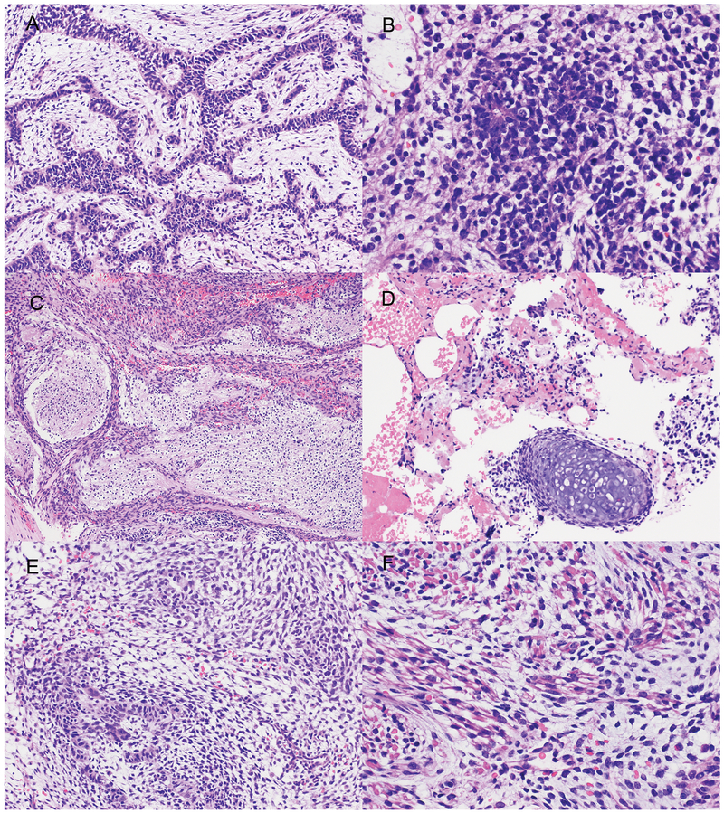
Figure 3.
Microscopic appearance of the multipatterned tumor from Patient 2.The tumor from Patient 2 was composed predominantly of immature neuroepithelium in epithelial cords (A), clusters with neuroblasts with rare true rosettes (B) and differentiating neurocytic areas in a neuropil background (C). Rare immature cartilage nodules were seen (D). Embryonal rhabdomyosarcomatous areas with skeletal muscle differentiation were also present (E, F). Hematoxylin and eosin: Original magnification x 200 (A, D, E); x 100 (C), x 400 (B, F).

