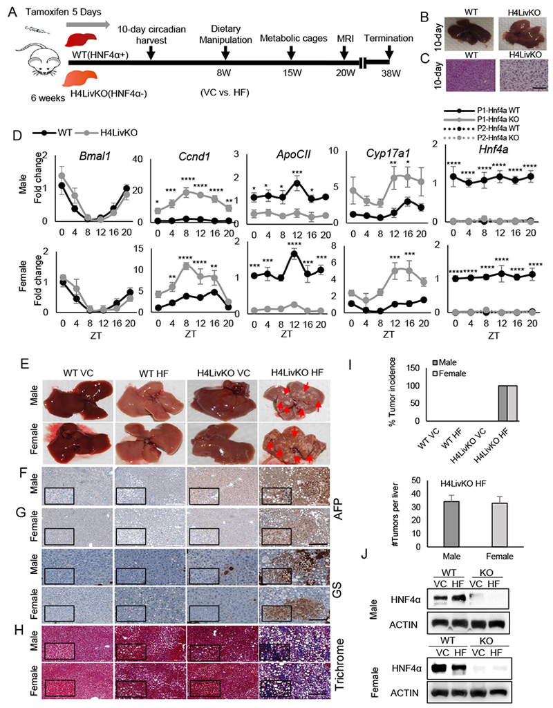
Figure 1. Loss of hepatic Hnf4a with HF feeding results in hepatocellular carcinoma (HCC) in male and female mice.

A. Experimental timeline. B. Livers of WT (Cre−) and H4LivKO (Cre+) mice, 10 days after initial tamoxifen injection. C. Hematoxylin and eosin (H&E) staining of WT and Hnf4a knockout (H4LivKO) liver (Scale bar=100 μm.) D. Hepatic gene expression in WT and H4LivKO mice 10 days after initial tamoxifen injection as measured by RT-PCR. E. Livers taken both sexes of WT and H4LivKO mice fed vivarium chow (VC) or high fat (HF) diet for 30 weeks. F-H. Staining of livers using antibodies to Alpha Fetoprotein (AFP), or glutamine synthetase (GS) (G) (Scale bar=200 μm.), and trichrome stain (H) (Scale bar=100 μm.) I. Percent tumor incidence and number of tumors per liver. (N = 8-16) J. Western blot of HNF4α in male and female H4LivKO livers under conditions of VC or HF feeding. Two-way ANOVA, Sidak’s multiple comparisons test, relative to WT, *P < 0.03, **P < 0.005, ***P < 0.0005, ****P < 0.0001. (N = 4-5) Error bars = SEM. (Table S5 for JTK_Cycle Rhythmicity Statistics.)
