
Figure 3. Loss of HNF4α followed by high fat feeding results in a permissive hepatic environment for HCC in male and female mice.
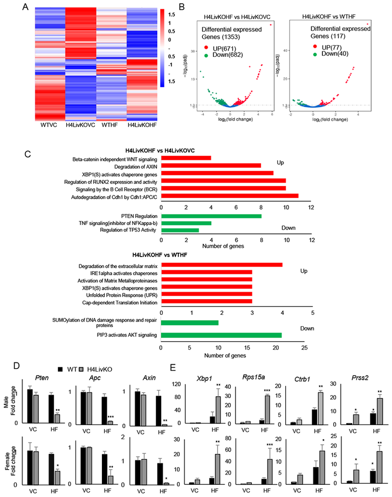
A. Heat maps generated from RNA-seq data reveal changes in gene expression between male WT vs. H4LivKO mice fed prolonged vivarium chow (VC) or high fat (HF) diet. B. Volcano plots reveal differentially expressed genes in H4LivKO fed HF diet (H4LivKOHF) or vivarium chow (H4LivKOVC), (left); or KOHF and WT mice fed HF diet for prolonged (30 weeks) (WTHF) (right) (N=3). C. Gene annotation reveals pathways altered in H4LivKOHF vs. H4LivKOVC (top) and H4LivKOHF vs. WTHF (bottom). D-E. RT-PCR reveals altered expression of genes involved in tumor suppression or inhibition of WNT/β catenin signaling (D) and genes involved in translation and protein folding (E) in WT and H4LivKO livers from animals fed VC or HF. Two-way ANOVA, Sidak’s multiple comparisons test (N=4-6) *P < 0.03, **P < 0.005, ***P < 0.0005, ****P < 0.0001.