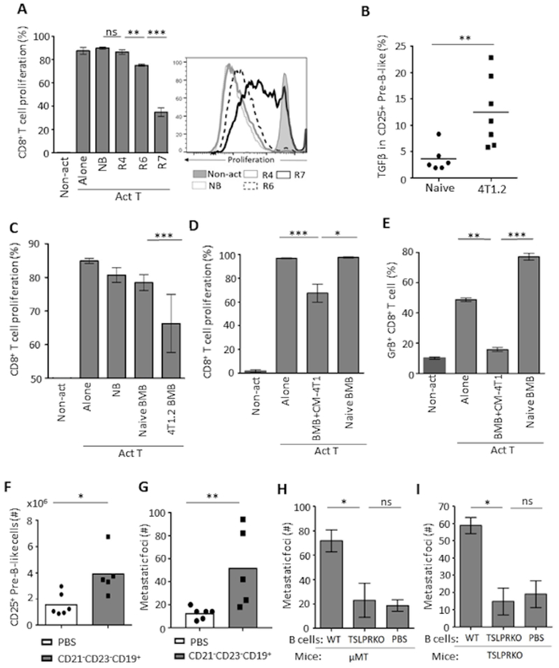
Figure 6. 4T1 cancer uses pre-B-like cells to generate tBregs (A–F) which promote metastasis (G–I).
Shown are representative data from in vitro CD8+ T cell suppression assay with pre-B-like cells from spleen (R6 and R7, A) or with BM B cells (both CD25+ and CD25− IgM− B-precursors, BMB, C) from mice with 4T1 cancer (4T1.2 BMB) or without cancer (NB). Control cells were FOB (R4) and BMB from naïve mice. Non-act is for unstimulated T cells and Act T is for T cells stimulated with anti-CD3e Ab (A) or with anti-CD3e/anti-CD28 Ab beads (C-E). Unlike circulating pre-B-like cells from cancer-free mice, pre-B-like cells from mice with 4T1 cancer express high levels of TGFβ (intracellular FACS staining result, B). To demonstrate that 4T1 cancer confers B-cell precursors with a regulatory activity, naïve mouse BM B-cell precursors (BMB) are cultured in CM-4T1 or in cRPMI for 7 days (D and E). Only CM-4T1-treated BMB suppressed proliferation (D) and granzyme B expression (E) in CD8+ T cells in in vitro suppression assay. In A, C, and D, proliferation is measured by the dilution of eFluor450 (a representative histogram, right panel in A) and depicted as mean ± SD of triplicate experiments reproduced at least twice (n=3-5). In F–G, BALB/c mice bearing 4T1.2 cancer (pretreated with 20 μg MK886 to eliminate endogenous tBregs) are i.v. injected with PBS or purified pre-B-like cells from spleen of syngeneic mice with 4T1.2 cancer (n=5). Shown are numbers of CD25+ pre-B-like cells in spleen (F) and metastatic foci in the lungs (G). Similarly, splenic B cells from WT or TSLPR KO mice with 4T1.2 cancer were i.v. transferred into μMT (H, n=4) and TSLPR KO mice with 4T1.2 cancer (I, n=4). Each symbol is for a single mouse (B, F, G) and Y-axis in G, H, I is for mean of metastatic foci ± SD in the lungs 34 days post tumor challenge. Results are reproduced at least twice. P-values were calculated with one-way ANOVA followed by Tukey test (A), Welch’s t-test (D, E), and Mann-Whitney Wilcoxon t-test (B, C, F–I).

