Figure 2.

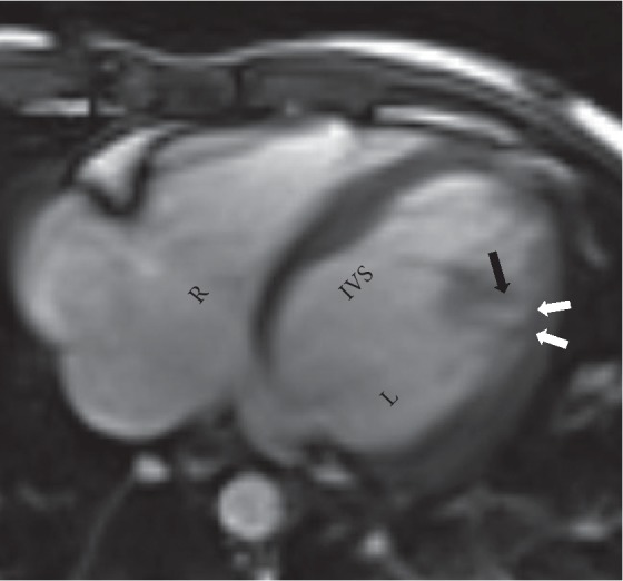
Axial steady-state free precession (SSFP) image depicting thinning of the compacted myocardium (white arrows) and prominent trabeculations along the lateral wall (black arrow) with noncompacted to compacted ratio >2.3.

Axial steady-state free precession (SSFP) image depicting thinning of the compacted myocardium (white arrows) and prominent trabeculations along the lateral wall (black arrow) with noncompacted to compacted ratio >2.3.