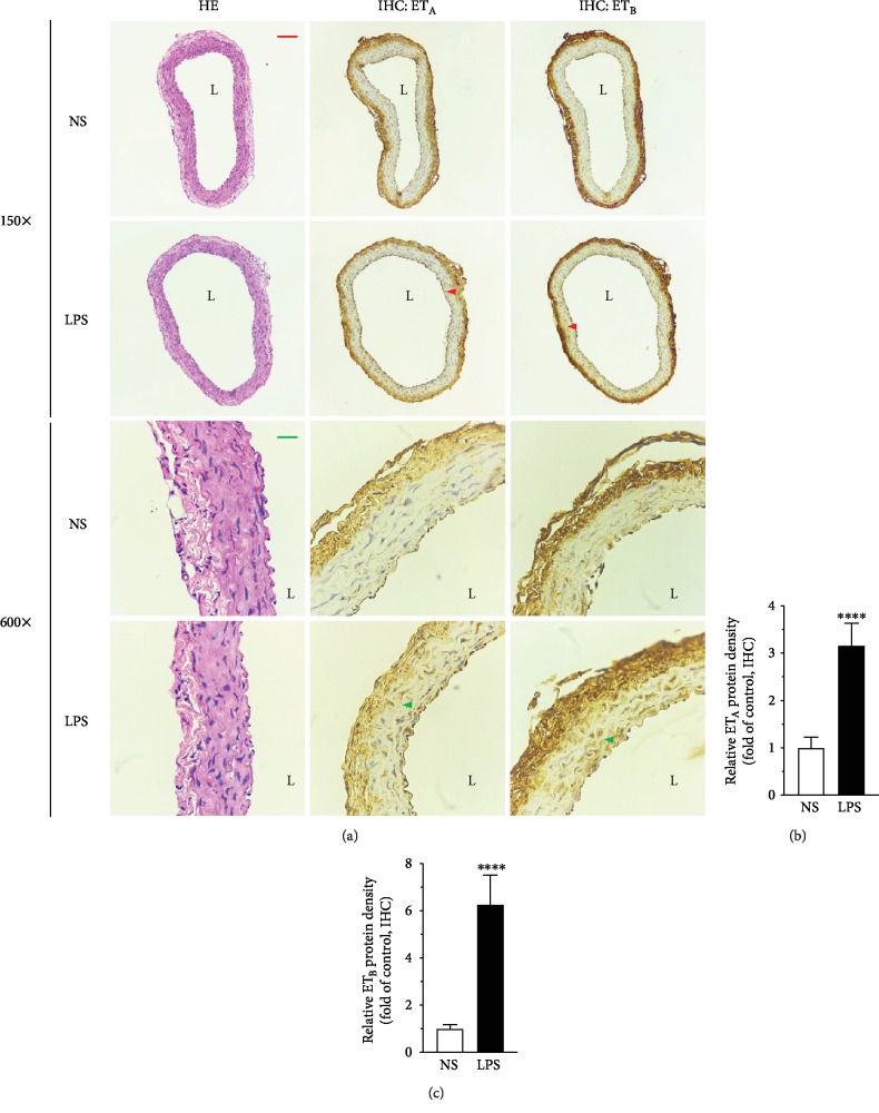
Figure 3.
Hematoxylin-eosin staining and immunohistochemical staining on paraffin-embedded tissue sections showed the morphology of the vessels and the upregulation of ETA and ETB after LPS treatment, respectively. Arteries (without endothelium) were taken from rats treated with NS or LPS (5 mg/kg body weight) for 6 hrs. The arrowhead points to the positive staining of ETA or ETB protein. L: lumen. The size bar corresponds to 200 μm (150x) or 50 μm (600x. (b, c) Semiquantitation of immunohistochemistry results by using ImageJ software. Data are expressed as mean ± SD. Unpaired Student's t-test, ∗∗∗∗P < 0.0001 versus NS, n = 10 sections (2 sections per animal) in each group.

