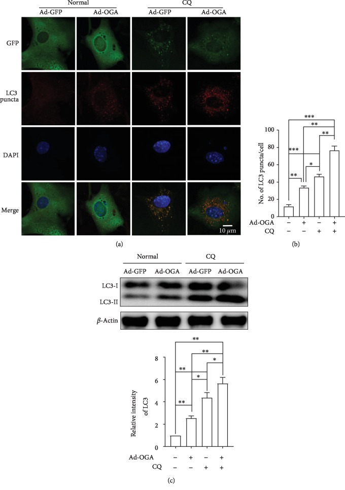
Figure 7.
OGA overexpression promotes autophagic flux in cortical astrocytes. (a) Astrocytes were transfected by Ad/eGFP and Ad-m-MGEA5/eGFP adenovirus for 48 h. Representative immunofluorescence was examined by anti-GFP and anti-LC3 antibodies. (b) Statistical analysis was calculated by counting the number of LC3 puncta-containing cells. Significance of different conditions was analyzed through ±SEM (∗p < 0.05, ∗∗p < 0.01, and ∗∗∗p < 0.001). (c) LC3 protein expressions were revealed by immunoblotting after overexpression of control and ad-OGA. Cells were treated with CQ before 2 h of cell lysates (n = 3, ∗p < 0.05, ∗∗p < 0.01).

