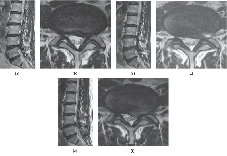
Figure 2.
Case of early recurrence after PELD. Preoperative MRI shows L4–5 disc herniation left paracentral and foraminal type (a, b). Immediate postoperative MRI image shows L4–5 left side disc removed and left nerve root decompressed (c, d). However, 4 month later, follow-up MRI shows L4–5 disc reherniation again at same operated site (e, f). According to scoring system, (1) age: 47 (2 point), (2) gender: male (0 point), (3) BMI: 28.3 kg/m2 (1 point), (4) disc degeneration scale: 3 scale (1 point), (5) Modic change (0 point), (6) combined HNP: 2 level (1 point), (7) disc herniation episode: first (0 point), (8) annulus preservation: minimal annulotomy (0 point), (9) approach: transforaminal outside-in (0 point) (10) disc location: paracentral (2 points) (11) herniated disc level: L4–5 (1 point), (12) disc height: 80–100% (0 point), (13) segmental dynamic motion: group 5 (0 point), (14) early ambulation: walking within 2 days (1 point), (15) spinal orthosis: no corset (1 point). Total score: 10 points (group C) .

