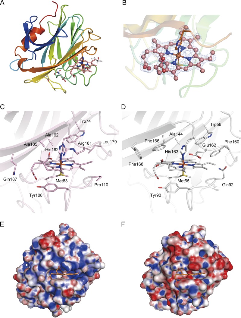
FIG 4.
Structure of the AA8 domain of CcPDH and comparison with the P. chrysosporium CDH AA8 domain. (A) Overall structure of the AA8 domain of CcPDH. Here, the Met83 residue, His182 residue, disulfide bond (Cys138-Cys141), 2-methyl-2,4-pentanediol molecule, acetate ion, and GlcNAc residue are shown as stick models. (B) Close-up view of heme b (pink) in the AA8 domain, with the 2Fo − Fc electron density map calculated at 1.8 Å and contoured at 1.5σ. (C, D) Heme b binding in the AA8 domain of CcPDH (C) and the PcCDH AA8 domain (D) (PDB accession number 1D7D). The surrounding amino acid residues are shown as stick models with labels. (E, F) Surface charge of the AA8 domain of CcPDH (E) and the PcCDH AA8 domain (F). Positively charged regions are colored blue, and negatively charged regions are colored red. Molecular surfaces were drawn by use of the PyMOL APBS plug-in and color coded from red (−10 kT) to blue (+10 kT).

